1ZWW

Crystal structure of endophilin-A1 BAR domain


Experimental Data Snapshot

  • Method: X-RAY DIFFRACTION
  • Resolution: 2.30 Å
  • R-Value Free: 0.287 
  • R-Value Work: 0.249 
  • R-Value Observed: 0.251 

wwPDB Validation   3D Report Full Report


This is version 1.3 of the entry. See complete history


Literature

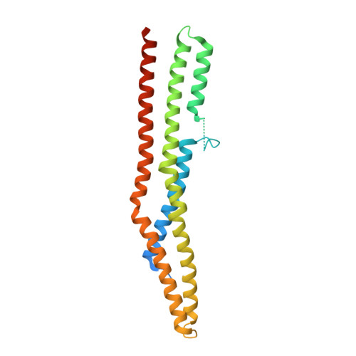
Crystal Structure of the Endophilin-A1 BAR Domain.

Weissenhorn, W.

(2005) J Mol Biol 351: 653-661

  • DOI: https://doi.org/10.1016/j.jmb.2005.06.013
  • Primary Citation of Related Structures:  
    1ZWW

  • PubMed Abstract: 

    Endophilin has been implicated in the retrieval of membrane via endocytosis of clathrin-coated vesicles, which is crucial for the maintenance of neurotransmitter exocytosis during stimulation; both exocytosis and endocytosis are regulated by intracellular calcium levels. Here, we present the 2.3 A crystal structure of the endophilin-A1 BAR domain, which has been suggested to function in inducing and sensing membrane curvature at the site of endocytosis. Endo-BAR folds into a crescent-shaped dimer composed of two elongated, three-helix bundles. Two additional domains of 30 residues each, inserted into helix 1 at the center of the concave side of the dimer, may interfere with the proposed mode of BAR domain membrane interaction. In addition, the dimer binds 11 divalent cadmium ions in the crystal mostly with typical Ca2+ co-ordination spheres. The endophilin-1A BAR domain thus constitutes a new variant of a BAR domain, and it may link endophilin-1A BAR function to calcium regulation of endocytosis.


  • Organizational Affiliation

    European Molecular Biology Laboratory (EMBL), 6 rue Jules Horowitz, 38042 Grenoble, France. weissen@embl-grenoble.fr


Macromolecules
Find similar proteins by:  (by identity cutoff)  |  3D Structure
Entity ID: 1
MoleculeChains Sequence LengthOrganismDetailsImage
SH3-containing GRB2-like protein 2
A, B
256Mus musculusMutation(s): 0 
Gene Names: Sh3gl2Sh3d2a
EC: 2.3.1
Membrane Entity: Yes 
UniProt & NIH Common Fund Data Resources
Find proteins for Q62420 (Mus musculus)
Explore Q62420 
Go to UniProtKB:  Q62420
IMPC:  MGI:700009
Entity Groups  
Sequence Clusters30% Identity50% Identity70% Identity90% Identity95% Identity100% Identity
UniProt GroupQ62420
Sequence Annotations
Expand
  • Reference Sequence
Small Molecules
Ligands 1 Unique
IDChains Name / Formula / InChI Key2D Diagram3D Interactions
CD
Query on CD

Download Ideal Coordinates CCD File 
C [auth A]
D [auth A]
E [auth A]
F [auth A]
G [auth A]
C [auth A],
D [auth A],
E [auth A],
F [auth A],
G [auth A],
H [auth A],
I [auth B],
J [auth B],
K [auth B],
L [auth B],
M [auth B]
CADMIUM ION
Cd
WLZRMCYVCSSEQC-UHFFFAOYSA-N
Experimental Data & Validation

Experimental Data

  • Method: X-RAY DIFFRACTION
  • Resolution: 2.30 Å
  • R-Value Free: 0.287 
  • R-Value Work: 0.249 
  • R-Value Observed: 0.251 
  • Space Group: C 2 2 21
Unit Cell:
Length ( Å )Angle ( ˚ )
a = 40.705α = 90
b = 121.651β = 90
c = 244.026γ = 90
Software Package:
Software NamePurpose
REFMACrefinement
PDB_EXTRACTdata extraction
DENZOdata reduction
SCALEPACKdata scaling
SOLVEphasing

Structure Validation

View Full Validation Report


Entry History 

Deposition Data

Revision History  (Full details and data files)

  • Version 1.0: 2005-08-02
    Type: Initial release
  • Version 1.1: 2008-04-30
    Changes: Version format compliance
  • Version 1.2: 2011-07-13
    Changes: Version format compliance
  • Version 1.3: 2024-02-14
    Changes: Data collection, Database references, Derived calculations