1ZWL

Structure of WrbA from Pseudomonas aeruginosa in complex with FMN


Experimental Data Snapshot

  • Method: X-RAY DIFFRACTION
  • Resolution: 2.80 Å
  • R-Value Free: 0.236 
  • R-Value Work: 0.197 
  • R-Value Observed: 0.199 

wwPDB Validation   3D Report Full Report


Ligand Structure Quality Assessment 


This is version 1.4 of the entry. See complete history


Literature

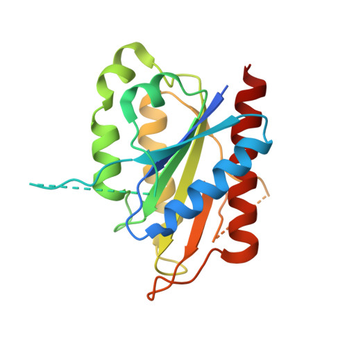
Crystal structures of the tryptophan repressor binding protein WrbA and complexes with flavin mononucleotide.

Gorman, J.Shapiro, L.

(2005) Protein Sci 14: 3004-3012

  • DOI: https://doi.org/10.1110/ps.051680805
  • Primary Citation of Related Structures:  
    1YDG, 1YRH, 1ZWK, 1ZWL

  • PubMed Abstract: 

    The tryptophan repressor binding protein WrbA binds to the tryptophan repressor protein TrpR. Although the biological role of WrbA remains unclear, it has been proposed to function in enhancing the stability of TrpR-DNA complexes. Sequence database analysis has identified WrbA as a founding member of a flavodoxin-like family of proteins. Here we present crystal structures of WrbA from Deinococcus radiodurans and Pseudomonas aeruginosa and their complexes with flavin mononucleotide. The protomer structure is similar to that of previously determined long-chain flavodoxins; however, each contains a conserved inserted region unique to the WrbA family. Interestingly, each WrbA protein forms a homotetramer with 222 symmetry, unique among flavodoxin-like proteins, in which each protomer binds one flavin mononucleotide cofactor molecule.


  • Organizational Affiliation

    Department of Biochemistry and Molecular Biophysics, Columbia University, 630 West 168th Street, Box 18, New York, NY 10032, USA.


Macromolecules
Find similar proteins by:  (by identity cutoff)  |  3D Structure
Entity ID: 1
MoleculeChains Sequence LengthOrganismDetailsImage
Trp repressor binding protein WrbA207Pseudomonas aeruginosaMutation(s): 0 
Gene Names: wrba
UniProt
Find proteins for Q9I509 (Pseudomonas aeruginosa (strain ATCC 15692 / DSM 22644 / CIP 104116 / JCM 14847 / LMG 12228 / 1C / PRS 101 / PAO1))
Explore Q9I509 
Go to UniProtKB:  Q9I509
Entity Groups  
Sequence Clusters30% Identity50% Identity70% Identity90% Identity95% Identity100% Identity
UniProt GroupQ9I509
Sequence Annotations
Expand
  • Reference Sequence
Small Molecules
Ligands 1 Unique
IDChains Name / Formula / InChI Key2D Diagram3D Interactions
FMN
Query on FMN

Download Ideal Coordinates CCD File 
B [auth A]FLAVIN MONONUCLEOTIDE
C17 H21 N4 O9 P
FVTCRASFADXXNN-SCRDCRAPSA-N
Experimental Data & Validation

Experimental Data

  • Method: X-RAY DIFFRACTION
  • Resolution: 2.80 Å
  • R-Value Free: 0.236 
  • R-Value Work: 0.197 
  • R-Value Observed: 0.199 
  • Space Group: P 42 2 2
Unit Cell:
Length ( Å )Angle ( ˚ )
a = 73.54α = 90
b = 73.54β = 90
c = 78.166γ = 90
Software Package:
Software NamePurpose
REFMACrefinement
DENZOdata reduction
SCALEPACKdata scaling
PHASERphasing

Structure Validation

View Full Validation Report


Ligand Structure Quality Assessment 


Entry History 

Deposition Data

Revision History  (Full details and data files)

  • Version 1.0: 2005-06-14
    Type: Initial release
  • Version 1.1: 2008-04-30
    Changes: Version format compliance
  • Version 1.2: 2011-07-13
    Changes: Derived calculations, Version format compliance
  • Version 1.3: 2021-02-03
    Changes: Database references, Derived calculations, Structure summary
  • Version 1.4: 2023-08-23
    Changes: Data collection, Database references, Refinement description