Crystal structures of reaction intermediates of L-2-haloacid dehalogenase and implications for the reaction mechanism.
Li, Y.F., Hata, Y., Fujii, T., Hisano, T., Nishihara, M., Kurihara, T., Esaki, N.(1998) J Biol Chem 273: 15035-15044
- PubMed: 9614112
- DOI: https://doi.org/10.1074/jbc.273.24.15035
- Primary Citation of Related Structures:
1ZRM, 1ZRN - PubMed Abstract:
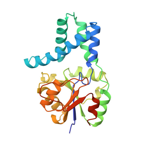
Crystal structures of L-2-haloacid dehalogenase from Pseudomonas sp. YL complexed with monochloroacetate, L-2-chlorobutyrate, L-2-chloro-3-methylbutyrate, or L-2-chloro-4-methylvalerate were determined at 1.83-, 2.0-, 2.2-, and 2.2-A resolutions, respectively, using the complex crystals prepared with the S175A mutant, which are isomorphous with those of the wild-type enzyme. These structures exhibit unique structural features that correspond to those of the reaction intermediates. In each case, the nucleophile Asp-10 is esterified with the dechlorinated moiety of the substrate. The substrate moieties in all but the monochloroacetate intermediate have a D-configuration at the C2 atom. The overall polypeptide fold of each of the intermediates is similar to that of the wild-type enzyme. However, it is clear that the Asp-10-Ser-20 region moves to the active site in all of the intermediates, and the Tyr-91-Asp-102 and Leu-117-Arg-135 regions make conformational changes in all but the monochloroacetate intermediates. Ser-118 is located near the carboxyl group of the substrate moiety; this residue probably serves as a binding residue for the substrate carboxyl group. The hydrophobic pocket, which is primarily composed of the Tyr-12, Gln-42, Leu-45, Phe-60, Lys-151, Asn-177, and Trp-179 side chains, exists around the alkyl group of the substrate moiety. This pocket may play an important role in stabilizing the alkyl group of the substrate moiety through hydrophobic interactions, and may also play a role in determining the stereospecificity of the enzyme. Moreover, a water molecule, which is absent in the substrate-free enzyme, is present in the vicinities of the carboxyl carbon of Asp-10 and the side chains of Asp-180, Asn-177, and Ala-175 in each intermediate. This water molecule may hydrolyze the ester intermediate and its substrate. These findings crystallographically demonstrate that the enzyme reaction proceeds through the formation of an ester intermediate with the enzyme's nucleophile Asp-10.
Organizational Affiliation:
Institute for Chemical Research, Kyoto University, Uji, Kyoto 611-0011, Japan.