Characterization of the metal ion binding helix-hairpin-helix motifs in human DNA polymerase beta by X-ray structural analysis.
Pelletier, H., Sawaya, M.R.(1996) Biochemistry 35: 12778-12787
- PubMed: 8841120
- DOI: https://doi.org/10.1021/bi960790i
- Primary Citation of Related Structures:
1NOM, 1ZQA, 1ZQB, 1ZQC, 1ZQD, 1ZQE, 1ZQF, 1ZQG, 1ZQH, 1ZQI, 1ZQJ, 1ZQK, 1ZQL, 1ZQM, 1ZQN, 1ZQO, 1ZQP, 1ZQQ, 1ZQR, 1ZQS, 1ZQU, 1ZQV, 1ZQW, 1ZQX, 1ZQY, 1ZQZ, 8ICA, 8ICB, 8ICC, 8ICE, 8ICF, 8ICG, 8ICH, 8ICI, 8ICZ, 9ICA, 9ICB, 9ICC, 9ICE, 9ICG, 9ICH, 9ICI, 9ICJ, 9ICL - PubMed Abstract:
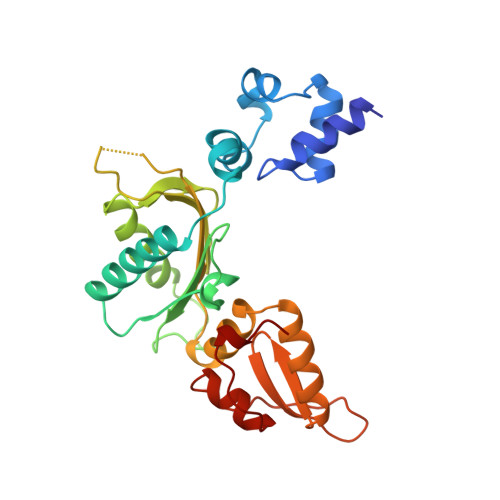
X-ray crystallographic studies have shown that DNA binding by human polymerase beta (pol beta) occurs primarily through two structurally and sequentially homologous helix-hairpin-helix (HhH) motifs, one in the fingers subdomain and the other in the 8-kDa domain [Pelletier, H., Sawaya, M. R., Wolfle, W., Wilson, S. H., & Kraut, J. (1996a) Biochemistry 35, 12742-12761]. In that DNA binding by each HhH motif is facilitated by a metal ion, we set out to determine the identity of the metal ion that most likely binds to the HhH motif in vivo. Crystal soaking experiments were performed on human pol beta-DNA cocrystals with Mg2+, Ca2+, Na+, and K+, the four most prevalent metal ions in the cell, and in each case a data set was collected and the resulting structure was refined. Under the conditions tested, the HhH motifs of pol beta have an affinity for these biologically prevalent metal ions in the order Mg2+ < Ca2+ < Na+ < K+, with K+ displaying the strongest binding. Crystals soaked in the presence of Tl+, a commonly used spectroscopic probe for K+, were too X-ray-sensitive to establish the binding behavior of Tl+, but soaking experiments with Ba2+ and Cs+ resulted in relatively stable crystals that gave evidence of metal ion binding in both HhH motifs, confirming that larger monovalent and divalent metal ions are capable of binding to the HhH metal sites. Although Mn2+, which has been categorized as a potent polymerase mutagen, binds to the HhH motifs with a greater affinity than Mg2+, Mn2+ does not bind to the HhH motifs in the presence of equimolar concentrations of Na+. These results suggest that in vivo, where Mn2+ is present only in trace amounts, Mn2+ probably does not have a large effect on DNA binding and may instead manifest a mutagenic effect on pol beta primarily by distorting nucleotide binding or by directly affecting the catalytic step [Pelletier, H., Sawaya, M. R., Wolfle, W., Wilson, S. H., & Kraut, J. (1996b) Biochemistry 35, 12762-12777]. Crystal soaking experiments with 31-kDa apoenzyme crystals show that, in the absence of DNA, the HhH motif in the fingers subdomain binds metal ions with either much lower occupancy or not at all, indicating that metal ion binding is dependent on the presence of the DNA substrate.
Organizational Affiliation:
Department of Chemistry and Biochemistry, University of California, San Diego, La Jolla 92093-0506, USA.