


Crystal structures of dialkylglycine decarboxylase bound with cesium ion and calcium ion
Liu, W., Toney, M.D.To be published.
Experimental Data Snapshot
Entity ID: 1 | |||||
|---|---|---|---|---|---|
| Molecule | Chains | Sequence Length | Organism | Details | Image |
| 2,2-dialkylglycine decarboxylase | 433 | Burkholderia cepacia | Mutation(s): 0 EC: 4.1.1.64 | 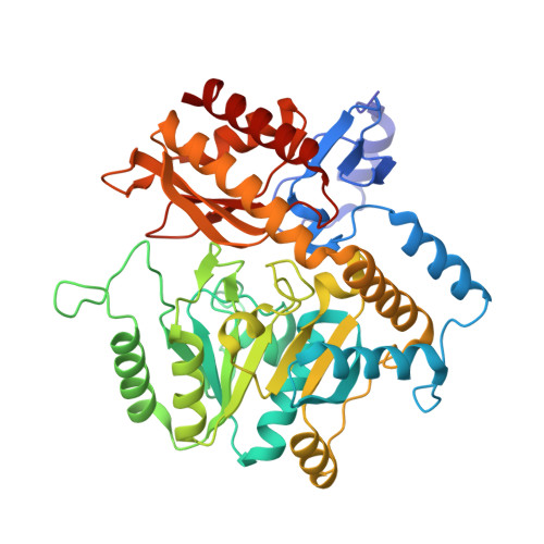 | |
UniProt | |||||
Find proteins for P16932 (Burkholderia cepacia) Explore P16932 Go to UniProtKB: P16932 | |||||
Entity Groups | |||||
| Sequence Clusters | 30% Identity50% Identity70% Identity90% Identity95% Identity100% Identity | ||||
| UniProt Group | P16932 | ||||
Sequence AnnotationsExpand | |||||
| |||||
| Ligands 4 Unique | |||||
|---|---|---|---|---|---|
| ID | Chains | Name / Formula / InChI Key | 2D Diagram | 3D Interactions | |
| PLP Query on PLP | D [auth A] | PYRIDOXAL-5'-PHOSPHATE C8 H10 N O6 P NGVDGCNFYWLIFO-UHFFFAOYSA-N |  | ||
| MES Query on MES | E [auth A] | 2-(N-MORPHOLINO)-ETHANESULFONIC ACID C6 H13 N O4 S SXGZJKUKBWWHRA-UHFFFAOYSA-N |  | ||
| CA Query on CA | C [auth A] | CALCIUM ION Ca BHPQYMZQTOCNFJ-UHFFFAOYSA-N |  | ||
| NA Query on NA | B [auth A] | SODIUM ION Na FKNQFGJONOIPTF-UHFFFAOYSA-N |  | ||
| Length ( Å ) | Angle ( ˚ ) |
|---|---|
| a = 150.553 | α = 90 |
| b = 150.553 | β = 90 |
| c = 84.703 | γ = 120 |
| Software Name | Purpose |
|---|---|
| PROTEUM PLUS | data collection |
| PROTEUM PLUS | data reduction |
| CNS | refinement |
| PROTEUM PLUS | data scaling |
| CNS | phasing |
RCSB PDB (citation) is hosted by
RCSB PDB is a member of the

