1ZMD

Crystal Structure of Human dihydrolipoamide dehydrogenase complexed to NADH


Experimental Data Snapshot

  • Method: X-RAY DIFFRACTION
  • Resolution: 2.08 Å
  • R-Value Free: 0.249 
  • R-Value Work: 0.221 
  • R-Value Observed: 0.221 

wwPDB Validation   3D Report Full Report


Ligand Structure Quality Assessment 


This is version 1.3 of the entry. See complete history


Literature

Crystal Structure of Human Dihydrolipoamide Dehydrogenase: NAD+/NADH Binding and the Structural Basis of Disease-causing Mutations

Brautigam, C.A.Chuang, J.L.Tomchick, D.R.Machius, M.Chuang, D.T.

(2005) J Mol Biol 350: 543-552

  • DOI: https://doi.org/10.1016/j.jmb.2005.05.014
  • Primary Citation of Related Structures:  
    1ZMC, 1ZMD

  • PubMed Abstract: 

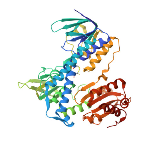
    Human dihydrolipoamide dehydrogenase (hE3) is an enzymatic component common to the mitochondrial alpha-ketoacid dehydrogenase and glycine decarboxylase complexes. Mutations to this homodimeric flavoprotein cause the often-fatal human disease known as E3 deficiency. To catalyze the oxidation of dihydrolipoamide, hE3 uses two molecules: non-covalently bound FAD and a transiently bound substrate, NAD+. To address the catalytic mechanism of hE3 and the structural basis for E3 deficiency, the crystal structures of hE3 in the presence of NAD+ or NADH have been determined at resolutions of 2.5A and 2.1A, respectively. Although the overall fold of the enzyme is similar to that of yeast E3, these two structures differ at two loops that protrude from the proteins and at their FAD-binding sites. The structure of oxidized hE3 with NAD+ bound demonstrates that the nicotinamide moiety is not proximal to the FAD. When NADH is present, however, the nicotinamide base stacks directly on the isoalloxazine ring system of the FAD. This is the first time that this mechanistically requisite conformation of NAD+ or NADH has been observed in E3 from any species. Because E3 structures were previously available only from unicellular organisms, speculations regarding the molecular mechanisms of E3 deficiency were based on homology models. The current hE3 structures show directly that the disease-causing mutations occur at three locations in the human enzyme: the dimer interface, the active site, and the FAD and NAD(+)-binding sites. The mechanisms by which these mutations impede the function of hE3 are discussed.


  • Organizational Affiliation

    Department of Biochemistry, The University of Texas, Southwestern Medical Center at Dallas, 5323 Harry Hines Boulevard, Dallas, TX 75390-9038, USA. chad.brautigam@utsouthwestern.edu


Macromolecules
Find similar proteins by:  (by identity cutoff)  |  3D Structure
Entity ID: 1
MoleculeChains Sequence LengthOrganismDetailsImage
Dihydrolipoyl dehydrogenase
A, B, C, D, E
A, B, C, D, E, F, G, H
474Homo sapiensMutation(s): 0 
Gene Names: DLDGCSLLADPHE3
EC: 1.8.1.4
UniProt & NIH Common Fund Data Resources
Find proteins for P09622 (Homo sapiens)
Explore P09622 
Go to UniProtKB:  P09622
PHAROS:  P09622
GTEx:  ENSG00000091140 
Entity Groups  
Sequence Clusters30% Identity50% Identity70% Identity90% Identity95% Identity100% Identity
UniProt GroupP09622
Sequence Annotations
Expand
  • Reference Sequence
Small Molecules
Ligands 3 Unique
IDChains Name / Formula / InChI Key2D Diagram3D Interactions
FAD
Query on FAD

Download Ideal Coordinates CCD File 
BB [auth H]
DA [auth D]
JA [auth E]
L [auth A]
PA [auth F]
BB [auth H],
DA [auth D],
JA [auth E],
L [auth A],
PA [auth F],
R [auth B],
VA [auth G],
X [auth C]
FLAVIN-ADENINE DINUCLEOTIDE
C27 H33 N9 O15 P2
VWWQXMAJTJZDQX-UYBVJOGSSA-N
NAI
Query on NAI

Download Ideal Coordinates CCD File 
CB [auth H]
EA [auth D]
KA [auth E]
M [auth A]
QA [auth F]
CB [auth H],
EA [auth D],
KA [auth E],
M [auth A],
QA [auth F],
S [auth B],
WA [auth G],
Y [auth C]
1,4-DIHYDRONICOTINAMIDE ADENINE DINUCLEOTIDE
C21 H29 N7 O14 P2
BOPGDPNILDQYTO-NNYOXOHSSA-N
SO4
Query on SO4

Download Ideal Coordinates CCD File 
AA [auth D]
AB [auth H]
BA [auth D]
CA [auth D]
FA [auth E]
AA [auth D],
AB [auth H],
BA [auth D],
CA [auth D],
FA [auth E],
GA [auth E],
HA [auth E],
I [auth A],
IA [auth E],
J [auth A],
K [auth A],
LA [auth F],
MA [auth F],
N [auth B],
NA [auth F],
O [auth B],
OA [auth F],
P [auth B],
Q [auth B],
RA [auth G],
SA [auth G],
T [auth C],
TA [auth G],
U [auth C],
UA [auth G],
V [auth C],
W [auth C],
XA [auth H],
YA [auth H],
Z [auth D],
ZA [auth H]
SULFATE ION
O4 S
QAOWNCQODCNURD-UHFFFAOYSA-L
Experimental Data & Validation

Experimental Data

  • Method: X-RAY DIFFRACTION
  • Resolution: 2.08 Å
  • R-Value Free: 0.249 
  • R-Value Work: 0.221 
  • R-Value Observed: 0.221 
  • Space Group: P 21 21 2
Unit Cell:
Length ( Å )Angle ( ˚ )
a = 175.871α = 90
b = 210.914β = 90
c = 127.096γ = 90
Software Package:
Software NamePurpose
CNSrefinement
HKL-2000data reduction
SCALEPACKdata scaling
AMoREphasing

Structure Validation

View Full Validation Report


Ligand Structure Quality Assessment 


Entry History 

Deposition Data

Revision History  (Full details and data files)

  • Version 1.0: 2005-06-28
    Type: Initial release
  • Version 1.1: 2008-04-30
    Changes: Version format compliance
  • Version 1.2: 2011-07-13
    Changes: Version format compliance
  • Version 1.3: 2023-08-23
    Changes: Data collection, Database references, Derived calculations, Refinement description