The apo and ternary complex structures of a chemotherapeutic target: human glycinamide ribonucleotide transformylase.
Dahms, T.E., Sainz, G., Giroux, E.L., Caperelli, C.A., Smith, J.L.(2005) Biochemistry 44: 9841-9850
- PubMed: 16026156
- DOI: https://doi.org/10.1021/bi050307g
- Primary Citation of Related Structures:
1ZLX, 1ZLY - PubMed Abstract:
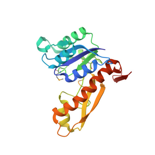
Glycinamide ribonucleotide transformylase (GART; 10-formyltetrahydrofolate:5'-phosphoribosylglycinamide formyltransferase, EC 2.1.2.2), an essential enzyme in de novo purine biosynthesis, has been a chemotherapeutic target for several decades. The three-dimensional structure of the GART domain from the human trifunctional enzyme has been solved by X-ray crystallography. Models of the apoenzyme, and a ternary complex with the 10-formyl-5,8-dideazafolate cosubstrate and a glycinamide ribonucleotide analogue, hydroxyacetamide ribonucleotide [alpha,beta-N-(hydroxyacetyl)-d-ribofuranosylamine], are reported to 2.2 and 2.07 A, respectively. The model of the apoenzyme represents the first structure of GART, from any source, with a completely unoccupied substrate and cosubstrate site, while the ternary complex is the first structure of the human GART domain that is bound at both the substrate and cosubstrate sites. A comparison of the two models therefore reveals subtle structural differences that reflect substrate and cosubstrate binding effects and implies roles for the invariant residues Gly 133, Gly 146, and His 137. Preactivation of the DDF formyl group appears to be key for catalysis, and structural flexibility of the active end of the substrate may facilitate nucleophilic attack. A change in pH, rather than folate binding, correlates with movement of the folate binding loop, whereas the phosphate binding loop position does not vary with pH. The electrostatic surface potentials of the human GART domain and Escherichia coli enzyme explain differences in the binding affinity of polyglutamylated folates, and these differences have implications to future chemotherapeutic agent design.
Organizational Affiliation:
Department of Biological Sciences, Purdue University, West Lafayette, Indiana 47907, USA.