1ZLH

Crystal structure of the tick carboxypeptidase inhibitor in complex with bovine carboxypeptidase A


Experimental Data Snapshot

  • Method: X-RAY DIFFRACTION
  • Resolution: 1.70 Å
  • R-Value Free: 0.185 
  • R-Value Work: 0.154 
  • R-Value Observed: 0.156 

wwPDB Validation   3D Report Full Report


This is version 1.3 of the entry. See complete history


Literature

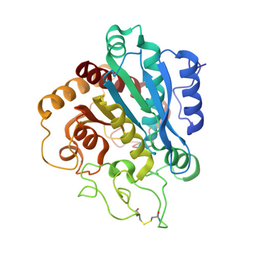
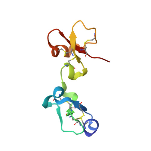
The Three-Dimensional Structures of Tick Carboxypeptidase Inhibitor in Complex with A/B Carboxypeptidases Reveal a Novel Double-headed Binding Mode

Arolas, J.L.Popowicz, G.M.Lorenzo, J.Sommerhoff, C.P.Huber, R.Aviles, F.X.Holak, T.A.

(2005) J Mol Biol 350: 489-498

  • DOI: https://doi.org/10.1016/j.jmb.2005.05.015
  • Primary Citation of Related Structures:  
    1ZLH, 1ZLI

  • PubMed Abstract: 

    The tick carboxypeptidase inhibitor (TCI) is a proteinaceous inhibitor of metallo-carboxypeptidases present in the blood-sucking tick Rhipicephalus bursa. The three-dimensional crystal structures of recombinant TCI bound to bovine carboxypeptidase A and to human carboxypeptidase B have been determined and refined at 1.7 A and at 2.0 A resolution, respectively. TCI consists of two domains that are structurally similar despite the low degree of sequence homology. The domains, each consisting of a short alpha-helix followed by a small twisted antiparallel beta-sheet, show a high level of structural homology to proteins of the beta-defensin-fold family. TCI anchors to the surface of mammalian carboxypeptidases in a double-headed manner not previously seen for carboxypeptidase inhibitors: the last three carboxy-terminal amino acid residues interact with the active site of the enzyme in a way that mimics substrate binding, and the N-terminal domain binds to an exosite distinct from the active-site groove. The structures of these complexes should prove valuable in the applications of TCI as a thrombolytic drug and as a basis for the design of novel bivalent carboxypeptidase inhibitors.


  • Organizational Affiliation

    Institut de Biotecnologia i Biomedicina and Departament de Bioquímica i Biologia Molecular, Universitat Autònoma de Barcelona, 08193 Bellaterra, Spain.


Macromolecules
Find similar proteins by:  (by identity cutoff)  |  3D Structure
Entity ID: 1
MoleculeChains Sequence LengthOrganismDetailsImage
Carboxypeptidase A1309Bos taurusMutation(s): 0 
EC: 3.4.17.1
UniProt
Find proteins for P00730 (Bos taurus)
Explore P00730 
Go to UniProtKB:  P00730
Entity Groups  
Sequence Clusters30% Identity50% Identity70% Identity90% Identity95% Identity100% Identity
UniProt GroupP00730
Sequence Annotations
Expand
  • Reference Sequence
Find similar proteins by:  (by identity cutoff)  |  3D Structure
Entity ID: 2
MoleculeChains Sequence LengthOrganismDetailsImage
carboxypeptidase inhibitor75Rhipicephalus bursaMutation(s): 0 
UniProt
Find proteins for Q5EPH2 (Rhipicephalus bursa)
Explore Q5EPH2 
Go to UniProtKB:  Q5EPH2
Entity Groups  
Sequence Clusters30% Identity50% Identity70% Identity90% Identity95% Identity100% Identity
UniProt GroupQ5EPH2
Sequence Annotations
Expand
  • Reference Sequence
Experimental Data & Validation

Experimental Data

  • Method: X-RAY DIFFRACTION
  • Resolution: 1.70 Å
  • R-Value Free: 0.185 
  • R-Value Work: 0.154 
  • R-Value Observed: 0.156 
  • Space Group: P 1 21 1
Unit Cell:
Length ( Å )Angle ( ˚ )
a = 47.07α = 90
b = 68.6β = 104.72
c = 57.89γ = 90
Software Package:
Software NamePurpose
REFMACrefinement
MAR345data collection
XDSdata scaling
MOLREPphasing

Structure Validation

View Full Validation Report


Entry History 

Deposition Data

Revision History  (Full details and data files)

  • Version 1.0: 2005-07-05
    Type: Initial release
  • Version 1.1: 2008-04-30
    Changes: Version format compliance
  • Version 1.2: 2011-07-13
    Changes: Version format compliance
  • Version 1.3: 2017-10-11
    Changes: Refinement description