1ZH0

Crystal Structure of L-3-(2-napthyl)alanine-tRNA synthetase in complex with L-3-(2-napthyl)alanine


Experimental Data Snapshot

  • Method: X-RAY DIFFRACTION
  • Resolution: 1.90 Å
  • R-Value Free: 0.275 
  • R-Value Work: 0.220 
  • R-Value Observed: 0.222 

wwPDB Validation   3D Report Full Report


Ligand Structure Quality Assessment 


This is version 1.4 of the entry. See complete history


Literature

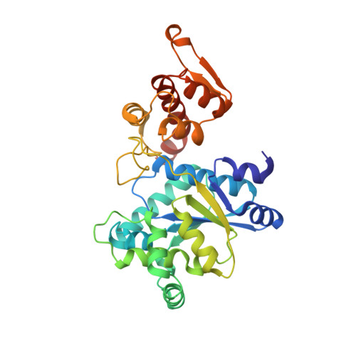
Structural plasticity of an aminoacyl-tRNA synthetase active site

Turner, J.M.Graziano, J.Spraggon, G.Schultz, P.G.

(2006) Proc Natl Acad Sci U S A 103: 6483-6488

  • DOI: https://doi.org/10.1073/pnas.0601756103
  • Primary Citation of Related Structures:  
    1ZH0, 2AG6

  • PubMed Abstract: 

    Recently, tRNA aminoacyl-tRNA synthetase pairs have been evolved that allow one to genetically encode a large array of unnatural amino acids in both prokaryotic and eukaryotic organisms. We have determined the crystal structures of two substrate-bound Methanococcus jannaschii tyrosyl aminoacyl-tRNA synthetases that charge the unnatural amino acids p-bromophenylalanine and 3-(2-naphthyl)alanine (NpAla). A comparison of these structures with the substrate-bound WT synthetase, as well as a mutant synthetase that charges p-acetylphenylalanine, shows that altered specificity is due to both side-chain and backbone rearrangements within the active site that modify hydrogen bonds and packing interactions with substrate, as well as disrupt the alpha8-helix, which spans the WT active site. The high degree of structural plasticity that is observed in these aminoacyl-tRNA synthetases is rarely found in other mutant enzymes with altered specificities and provides an explanation for the surprising adaptability of the genetic code to novel amino acids.


  • Organizational Affiliation

    Department of Chemistry and The Skaggs Institute for Chemical Biology, The Scripps Research Institute, 10550 North Torrey Pines Road, La Jolla, CA 92037, USA.


Macromolecules
Find similar proteins by:  (by identity cutoff)  |  3D Structure
Entity ID: 1
MoleculeChains Sequence LengthOrganismDetailsImage
Tyrosyl-tRNA synthetase314Methanocaldococcus jannaschiiMutation(s): 6 
EC: 6.1.1.1
UniProt
Find proteins for Q57834 (Methanocaldococcus jannaschii (strain ATCC 43067 / DSM 2661 / JAL-1 / JCM 10045 / NBRC 100440))
Explore Q57834 
Go to UniProtKB:  Q57834
Entity Groups  
Sequence Clusters30% Identity50% Identity70% Identity90% Identity95% Identity100% Identity
UniProt GroupQ57834
Sequence Annotations
Expand
  • Reference Sequence
Small Molecules
Ligands 2 Unique
IDChains Name / Formula / InChI Key2D Diagram3D Interactions
NAL
Query on NAL

Download Ideal Coordinates CCD File 
B [auth A]BETA-(2-NAPHTHYL)-ALANINE
C13 H13 N O2
JPZXHKDZASGCLU-LBPRGKRZSA-N
TRS
Query on TRS

Download Ideal Coordinates CCD File 
C [auth A]2-AMINO-2-HYDROXYMETHYL-PROPANE-1,3-DIOL
C4 H12 N O3
LENZDBCJOHFCAS-UHFFFAOYSA-O
Experimental Data & Validation

Experimental Data

  • Method: X-RAY DIFFRACTION
  • Resolution: 1.90 Å
  • R-Value Free: 0.275 
  • R-Value Work: 0.220 
  • R-Value Observed: 0.222 
  • Space Group: P 43 21 2
Unit Cell:
Length ( Å )Angle ( ˚ )
a = 102.995α = 90
b = 102.995β = 90
c = 71.441γ = 90
Software Package:
Software NamePurpose
DENZOdata reduction
SCALEPACKdata scaling
MOLREPphasing
REFMACrefinement
PDB_EXTRACTdata extraction
HKL-2000data reduction

Structure Validation

View Full Validation Report


Ligand Structure Quality Assessment 


Entry History 

Deposition Data

Revision History  (Full details and data files)

  • Version 1.0: 2006-04-04
    Type: Initial release
  • Version 1.1: 2008-04-02
    Changes: Version format compliance
  • Version 1.2: 2011-07-13
    Changes: Advisory, Derived calculations, Version format compliance
  • Version 1.3: 2021-10-20
    Changes: Database references, Derived calculations
  • Version 1.4: 2024-02-14
    Changes: Data collection