1ZCM

Human calpain protease core inhibited by ZLLYCH2F


Experimental Data Snapshot

  • Method: X-RAY DIFFRACTION
  • Resolution: 2.00 Å
  • R-Value Free: 0.223 
  • R-Value Work: 0.182 
  • R-Value Observed: 0.182 

wwPDB Validation   3D Report Full Report


Ligand Structure Quality Assessment 


This is version 1.5 of the entry. See complete history


Literature

Molecular mode of action of a covalently inhibiting peptidomimetic on the human calpain protease core

Li, Q.Hanzlik, R.P.Weaver, R.F.Schonbrunn, E.

(2006) Biochemistry 45: 701-708

  • DOI: https://doi.org/10.1021/bi052077b
  • Primary Citation of Related Structures:  
    1ZCM

  • PubMed Abstract: 

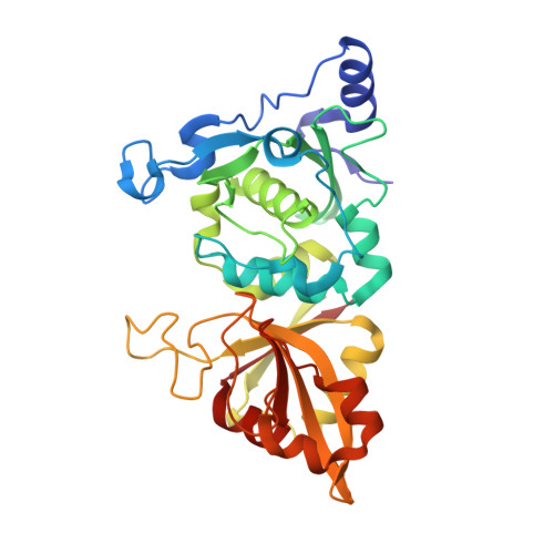
    Calpain is a nearly ubiquitous Ca2+-activated proteolytic enzyme whose precise physiological function is unknown. However, aberrant Ca2+ homeostasis in the course of cellular injuries and certain diseases of the CNS appears to activate calpain, in turn promoting the degradation of key cytoskeletal and membrane proteins. Hyperactive calpain has also been implicated in various aging phenomena and diseases of late life. Therefore, calpain is considered a potential therapeutic target in preventing degenerations of many kinds. Despite its potential medicinal importance, known structural information about mu-calpain is limited to that from the rat enzyme. We have determined the crystal structure of the human mu-calpain protease core (hmuI-II) containing a Gly213Ala mutation and covalently inactivated by a peptidomimetic (ZLLYCH2F) at 2.0 A resolution. The methylene carbon of the inhibitor is bound to Cys115. Additional hydrogen bonding and hydrophobic interactions between active site residues and the inhibitor, including an intermolecular antiparallel beta-sheet arrangement characteristically observed with members of the papain family of cysteine proteinases, help to stabilize the complex and orient the inhibitor. The terminal ZL portion of the inhibitor is solvent-exposed and highly flexible, and is thus not involved in specific binding interactions with the enzyme. Two large enzyme regions flanking the active site are highly flexible; they may be important in recognition of calpain's natural protein substrates and in positioning them for catalysis. The implications for drug design are discussed.


  • Organizational Affiliation

    Department of Medicinal Chemistry, University of Kansas, Lawrence, Kansas 66045, USA.


Macromolecules
Find similar proteins by:  (by identity cutoff)  |  3D Structure
Entity ID: 1
MoleculeChains Sequence LengthOrganismDetailsImage
Calpain 1, large [catalytic] subunit321Homo sapiensMutation(s): 1 
Gene Names: CAPN1CANPL1
EC: 3.4.22.52
UniProt & NIH Common Fund Data Resources
Find proteins for P07384 (Homo sapiens)
Explore P07384 
Go to UniProtKB:  P07384
PHAROS:  P07384
GTEx:  ENSG00000014216 
Entity Groups  
Sequence Clusters30% Identity50% Identity70% Identity90% Identity95% Identity100% Identity
UniProt GroupP07384
Sequence Annotations
Expand
  • Reference Sequence
Small Molecules
Ligands 2 Unique
IDChains Name / Formula / InChI Key2D Diagram3D Interactions
C1N
Query on C1N

Download Ideal Coordinates CCD File 
D [auth A]N-[(BENZYLOXY)CARBONYL]LEUCYL-N~1~-[3-FLUORO-1-(4-HYDROXYBENZYL)-2-OXOPROPYL]LEUCINAMIDE
C30 H40 F N3 O6
JCRSHQCFRMCMOC-GSDHBNRESA-N
CA
Query on CA

Download Ideal Coordinates CCD File 
B [auth A],
C [auth A]
CALCIUM ION
Ca
BHPQYMZQTOCNFJ-UHFFFAOYSA-N
Experimental Data & Validation

Experimental Data

  • Method: X-RAY DIFFRACTION
  • Resolution: 2.00 Å
  • R-Value Free: 0.223 
  • R-Value Work: 0.182 
  • R-Value Observed: 0.182 
  • Space Group: P 21 21 21
Unit Cell:
Length ( Å )Angle ( ˚ )
a = 50.44α = 90
b = 62.74β = 90
c = 99.26γ = 90
Software Package:
Software NamePurpose
XDSdata reduction
CNSrefinement
XDSdata scaling
CNSphasing

Structure Validation

View Full Validation Report


Ligand Structure Quality Assessment 


Entry History 

Deposition Data

Revision History  (Full details and data files)

  • Version 1.0: 2006-01-31
    Type: Initial release
  • Version 1.1: 2008-04-01
    Changes: Version format compliance
  • Version 1.2: 2011-07-13
    Changes: Version format compliance
  • Version 1.3: 2017-10-11
    Changes: Refinement description
  • Version 1.4: 2021-10-20
    Changes: Advisory, Database references, Derived calculations
  • Version 1.5: 2023-08-23
    Changes: Data collection, Refinement description