1ZAU

Adenylation domain of NAD+ dependent DNA ligase from M.tuberculosis


Experimental Data Snapshot

  • Method: X-RAY DIFFRACTION
  • Resolution: 3.15 Å
  • R-Value Free: 0.314 
  • R-Value Work: 0.253 
  • R-Value Observed: 0.253 

wwPDB Validation   3D Report Full Report


Ligand Structure Quality Assessment 


This is version 1.4 of the entry. See complete history


Literature

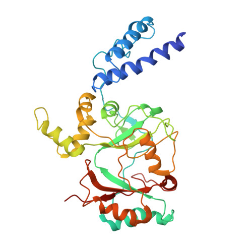
NAD+-dependent DNA Ligase (Rv3014c) from Mycobacterium tuberculosis: CRYSTAL STRUCTURE OF THE ADENYLATION DOMAIN AND IDENTIFICATION OF NOVEL INHIBITORS

Srivastava, S.K.Tripathi, R.P.Ramachandran, R.

(2005) J Biol Chem 280: 30273-30281

  • DOI: https://doi.org/10.1074/jbc.M503780200
  • Primary Citation of Related Structures:  
    1ZAU

  • PubMed Abstract: 

    DNA ligases utilize either ATP or NAD+ as cofactors to catalyze the formation of phosphodiester bonds in nicked DNA. Those utilizing NAD+ are attractive drug targets because of the unique cofactor requirement for ligase activity. We report here the crystal structure of the adenylation domain of the Mycobacterium tuberculosis NAD+-dependent ligase with bound AMP. The adenosine nucleoside moiety of AMP adopts a syn-conformation. The structure also captures a new spatial disposition between the two subdomains of the adenylation domain. Based on the crystal structure and an in-house compound library, we have identified a novel class of inhibitors for the enzyme using in silico docking calculations. The glycosyl ureide-based inhibitors were able to distinguish between NAD+- and ATP-dependent ligases as evidenced by in vitro assays using T4 ligase and human DNA ligase I. Moreover, assays involving an Escherichia coli strain harboring a temperature-sensitive ligase mutant and a ligase-deficient Salmonella typhimurium strain suggested that the bactericidal activity of the inhibitors is due to inhibition of the essential ligase enzyme. The results can be used as the basis for rational design of novel antibacterial agents.


  • Organizational Affiliation

    Division Molecular and Structural Biology, Central Drug Research Institute, Chattar Manzil, Mahatma Gandhi Marg, Lucknow-226001, India.


Macromolecules
Find similar proteins by:  (by identity cutoff)  |  3D Structure
Entity ID: 1
MoleculeChains Sequence LengthOrganismDetailsImage
DNA ligase328Mycobacterium tuberculosisMutation(s): 0 
Gene Names: ligAlig
EC: 6.5.1.2
UniProt
Find proteins for P9WNV1 (Mycobacterium tuberculosis (strain ATCC 25618 / H37Rv))
Explore P9WNV1 
Go to UniProtKB:  P9WNV1
Entity Groups  
Sequence Clusters30% Identity50% Identity70% Identity90% Identity95% Identity100% Identity
UniProt GroupP9WNV1
Sequence Annotations
Expand
  • Reference Sequence
Small Molecules
Ligands 1 Unique
IDChains Name / Formula / InChI Key2D Diagram3D Interactions
AMP
Query on AMP

Download Ideal Coordinates CCD File 
B [auth A]ADENOSINE MONOPHOSPHATE
C10 H14 N5 O7 P
UDMBCSSLTHHNCD-KQYNXXCUSA-N
Experimental Data & Validation

Experimental Data

  • Method: X-RAY DIFFRACTION
  • Resolution: 3.15 Å
  • R-Value Free: 0.314 
  • R-Value Work: 0.253 
  • R-Value Observed: 0.253 
  • Space Group: P 61 2 2
Unit Cell:
Length ( Å )Angle ( ˚ )
a = 95.8α = 90
b = 95.8β = 90
c = 208.4γ = 120
Software Package:
Software NamePurpose
MAR345data collection
SCALEPACKdata scaling
AMoREphasing
X-PLORrefinement

Structure Validation

View Full Validation Report


Ligand Structure Quality Assessment 


Entry History 

Deposition Data

Revision History  (Full details and data files)

  • Version 1.0: 2005-07-05
    Type: Initial release
  • Version 1.1: 2008-04-30
    Changes: Version format compliance
  • Version 1.2: 2011-07-13
    Changes: Version format compliance
  • Version 1.3: 2017-10-11
    Changes: Refinement description
  • Version 1.4: 2024-02-14
    Changes: Data collection, Database references, Derived calculations