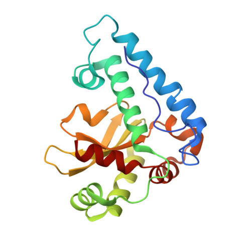
The Crucial Importance of Chemistry in the Structure-Function Link: Manipulating Hydrogen Bonding in Iron-Containing Superoxide Dismutase.
Yikilmaz, E., Rodgers, D.W., Miller, A.F.(2006) Biochemistry 45: 1151-1161
- PubMed: 16430211
- DOI: https://doi.org/10.1021/bi051495d
- Primary Citation of Related Structures:
1ZA5, 2BKB - PubMed Abstract:
Fe-containing superoxide dismutase's active site Fe is coordinated by a solvent molecule, whose protonation state is coupled to the Fe oxidation state. Thus, we have proposed that H-bonding between glutamine 69 and this solvent molecule can strongly influence the redox activity of the Fe in superoxide dismutase (SOD). We show here that mutation of this Gln to His subtly alters the active site structure but preserves 30% activity. In contrast, mutation to Glu otherwise preserves the active site structure but inactivates the enzyme. Thus, enzyme function correlates not with atom positions but with residue identity (chemistry), in this case. We observe strong destabilization of the Q69E-FeSOD oxidized state relative to the reduced state and intermediate destabilization of oxidized Q69H-FeSOD. Indeed, redox titrations indicate that mutation of Gln69 to His increases the reduction potential by 240 mV, whereas mutation to Glu appears to increase it by more than 660 mV. We find that this suffices to explain the mutants' loss of activity, although additional factors may also contribute. The strongly elevated reduction potential of Q69E-FeSOD may reflect reorganization of the active site H-bonding network, including possible reversal of the polarity of the key H-bond between residue 69 and coordinated solvent.
Organizational Affiliation:
Department of Chemistry, University of Kentucky, Lexington, Kentucky 40506, USA.