1Z9O

1.9 Angstrom Crystal Structure of the Rat VAP-A MSP Homology Domain in Complex with the Rat ORP1 FFAT Motif


Experimental Data Snapshot

  • Method: X-RAY DIFFRACTION
  • Resolution: 1.90 Å
  • R-Value Free: 0.261 
  • R-Value Work: 0.215 
  • R-Value Observed: 0.219 

wwPDB Validation   3D Report Full Report


This is version 1.3 of the entry. See complete history


Literature

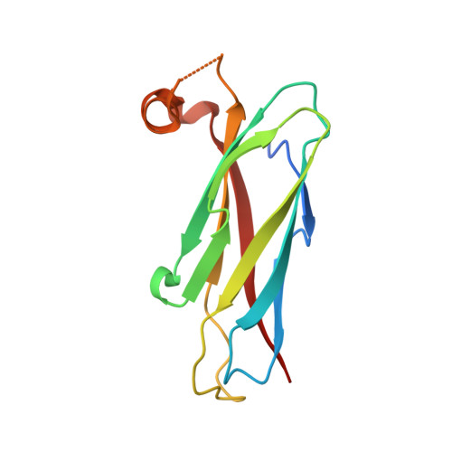
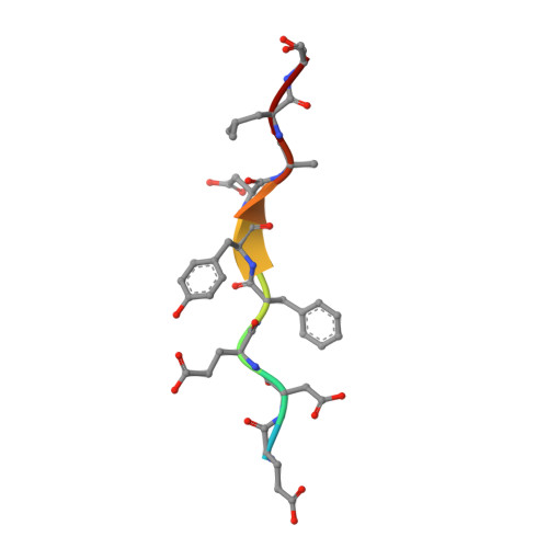
Structural basis of FFAT motif-mediated ER targeting

Kaiser, S.E.Brickner, J.H.Reilein, A.R.Fenn, T.D.Walter, P.Brunger, A.T.

(2005) Structure 13: 1035-1045

  • DOI: https://doi.org/10.1016/j.str.2005.04.010
  • Primary Citation of Related Structures:  
    1Z9L, 1Z9O

  • PubMed Abstract: 

    The FFAT motif is a targeting signal responsible for localizing a number of proteins to the cytosolic surface of the endoplasmic reticulum (ER) and to the nuclear membrane. FFAT motifs bind to members of the highly conserved VAP protein family, which are tethered to the cytoplasmic face of the ER by a C-terminal transmembrane domain. We have solved crystal structures of the rat VAP-A MSP homology domain alone and in complex with an FFAT motif. The co-crystal structure was used to design a VAP mutant that disrupts rat and yeast VAP-FFAT interactions in vitro. The FFAT binding-defective mutant also blocked function of the VAP homolog Scs2p in yeast. Finally, overexpression of the FFAT binding-defective VAP in COS7 cells dramatically altered ER morphology. Our data establish the structural basis of FFAT-mediated ER targeting and suggest that FFAT-targeted proteins play an important role in determining ER morphology.


  • Organizational Affiliation

    Department of Molecular and Cellular Physiology, Stanford University, Stanford, CA 94305, USA.


Macromolecules
Find similar proteins by:  (by identity cutoff)  |  3D Structure
Entity ID: 1
MoleculeChains Sequence LengthOrganismDetailsImage
Vesicle-associated membrane protein-associated protein A
A, B, C, D, E
A, B, C, D, E, F
128Rattus norvegicusMutation(s): 0 
Gene Names: VapaVap33
UniProt
Find proteins for Q9Z270 (Rattus norvegicus)
Explore Q9Z270 
Go to UniProtKB:  Q9Z270
Entity Groups  
Sequence Clusters30% Identity50% Identity70% Identity90% Identity95% Identity100% Identity
UniProt GroupQ9Z270
Sequence Annotations
Expand
  • Reference Sequence

Find similar proteins by:  Sequence   |   3D Structure  

Entity ID: 2
MoleculeChains Sequence LengthOrganismDetailsImage
Oxysterol binding protein
G, H, I, J, K
G, H, I, J, K, L
10N/AMutation(s): 0 
UniProt
Find proteins for Q8K4M9 (Rattus norvegicus)
Explore Q8K4M9 
Go to UniProtKB:  Q8K4M9
Entity Groups  
Sequence Clusters30% Identity50% Identity70% Identity90% Identity95% Identity100% Identity
UniProt GroupQ8K4M9
Sequence Annotations
Expand
  • Reference Sequence
Experimental Data & Validation

Experimental Data

  • Method: X-RAY DIFFRACTION
  • Resolution: 1.90 Å
  • R-Value Free: 0.261 
  • R-Value Work: 0.215 
  • R-Value Observed: 0.219 
  • Space Group: P 1
Unit Cell:
Length ( Å )Angle ( ˚ )
a = 50.048α = 90.01
b = 50.033β = 90
c = 90.287γ = 60.03
Software Package:
Software NamePurpose
REFMACrefinement
MOSFLMdata reduction
CCP4data scaling
CNSphasing

Structure Validation

View Full Validation Report


Entry History 

Deposition Data

Revision History  (Full details and data files)

  • Version 1.0: 2005-07-19
    Type: Initial release
  • Version 1.1: 2008-04-30
    Changes: Version format compliance
  • Version 1.2: 2011-07-13
    Changes: Advisory, Version format compliance
  • Version 1.3: 2023-08-23
    Changes: Data collection, Database references, Refinement description