1Z93

Human Carbonic Anhydrase III:Structural and Kinetic study of Catalysis and Proton Transfer.


Experimental Data Snapshot

  • Method: X-RAY DIFFRACTION
  • Resolution: 2.10 Å
  • R-Value Free: 0.258 
  • R-Value Work: 0.205 

wwPDB Validation   3D Report Full Report


This is version 1.4 of the entry. See complete history


Literature

Human Carbonic Anhydrase III: Structural and Kinetic Study of Catalysis and Proton Transfer

Duda, D.M.Tu, C.Fisher, S.Z.An, H.Yoshioka, C.Govindasamy, L.Laipis, P.J.Agbandje-McKenna, M.Silverman, D.N.McKenna, R.

(2005) Biochemistry 44: 10046-10053

  • DOI: https://doi.org/10.1021/bi050610h
  • Primary Citation of Related Structures:  
    1Z93, 1Z97

  • PubMed Abstract: 

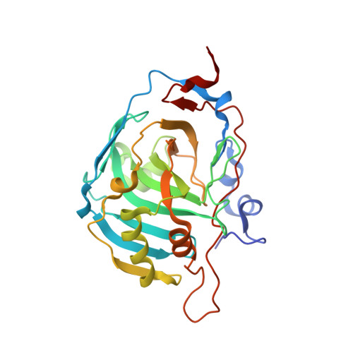
    The residue phenylalanine 198 (Phe 198) is a prominent cause of the lower activity of human carbonic anhydrase III (HCA III) compared with HCA II and other isozymes which have leucine at this site. We report the crystal structures of HCA III and the site-directed mutant F198L HCA III, both at 2.1 A resolution, and the enhancement of catalytic activity by exogenous proton donors containing imidazole rings. Both enzymes had a hexahistidine extension at the carboxy-terminal end, used to aid in purification, that was ordered in the crystal structures bound in the active site cavity of an adjacent symmetry-related enzyme. This observation allowed us to comment on a number of possible binding sites for imidazole and derivatives as exogenous proton donors/acceptors in catalysis by HCA III. Kinetic and structural evidence indicates that the phenyl side chain of Phe 198 in HCA III, about 5 A from the zinc, is a steric constriction in the active site, may cause altered interactions at the zinc-bound solvent, and is a binding site for the activation of catalysis by histidylhistidine. This suggests that sites of activation of the proton-transfer pathway in carbonic anhydrase are closer to the zinc than considered in previous studies.


  • Organizational Affiliation

    Departments of Biochemistry and Molecular Biology, College of Medicine, University of Florida, Gainesville, Florida 32610, USA.


Macromolecules
Find similar proteins by:  (by identity cutoff)  |  3D Structure
Entity ID: 1
MoleculeChains Sequence LengthOrganismDetailsImage
Carbonic anhydrase III266Homo sapiensMutation(s): 2 
Gene Names: CA3
EC: 4.2.1.1
UniProt & NIH Common Fund Data Resources
Find proteins for P07451 (Homo sapiens)
Explore P07451 
Go to UniProtKB:  P07451
PHAROS:  P07451
GTEx:  ENSG00000164879 
Entity Groups  
Sequence Clusters30% Identity50% Identity70% Identity90% Identity95% Identity100% Identity
UniProt GroupP07451
Sequence Annotations
Expand
  • Reference Sequence
Small Molecules
Ligands 1 Unique
IDChains Name / Formula / InChI Key2D Diagram3D Interactions
ZN
Query on ZN

Download Ideal Coordinates CCD File 
B [auth A]ZINC ION
Zn
PTFCDOFLOPIGGS-UHFFFAOYSA-N
Experimental Data & Validation

Experimental Data

  • Method: X-RAY DIFFRACTION
  • Resolution: 2.10 Å
  • R-Value Free: 0.258 
  • R-Value Work: 0.205 
  • Space Group: P 65
Unit Cell:
Length ( Å )Angle ( ˚ )
a = 44.7α = 90
b = 44.7β = 90
c = 222.557γ = 120
Software Package:
Software NamePurpose
DENZOdata reduction
SCALEPACKdata scaling
CNSrefinement
CNSphasing

Structure Validation

View Full Validation Report


Entry History 

Deposition Data

Revision History  (Full details and data files)

  • Version 1.0: 2005-08-09
    Type: Initial release
  • Version 1.1: 2008-04-30
    Changes: Version format compliance
  • Version 1.2: 2011-07-13
    Changes: Version format compliance
  • Version 1.3: 2021-10-20
    Changes: Database references, Derived calculations
  • Version 1.4: 2023-08-23
    Changes: Data collection, Refinement description