1Z3E

Crystal Structure of Spx in Complex with the C-terminal Domain of the RNA Polymerase Alpha Subunit


Experimental Data Snapshot

  • Method: X-RAY DIFFRACTION
  • Resolution: 1.50 Å
  • R-Value Free: 0.220 
  • R-Value Work: 0.196 
  • R-Value Observed: 0.196 

wwPDB Validation   3D Report Full Report


This is version 1.3 of the entry. See complete history


Literature

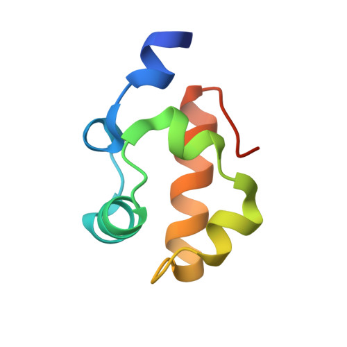
Crystal structure of the Bacillus subtilis anti-alpha, global transcriptional regulator, Spx, in complex with the {alpha} C-terminal domain of RNA polymerase

Newberry, K.J.Nakano, S.Zuber, P.Brennan, R.G.

(2005) Proc Natl Acad Sci U S A 102: 15839-15844

  • DOI: https://doi.org/10.1073/pnas.0506592102
  • Primary Citation of Related Structures:  
    1Z3E

  • PubMed Abstract: 

    Spx, a global transcription regulator in Bacillus subtilis, interacts with the C-terminal domain of the alpha subunit (alphaCTD) of RNA polymerase to control gene expression under conditions of disulfide stress, which is sensed by disulfide bond formation between Spx residues C10 and C13. Here, we describe the crystal structure of the B. subtilis alphaCTD bound to oxidized Spx. Analysis of the complex reveals interactions between three regions of "anti-alpha" Spx and helix alpha1 and the "261" determinant of alphaCTD. The former contact could disrupt the interaction between alphaCTD and activator proteins or alter the DNA-bound conformation of alphaCTD, thereby repressing activator-stimulated transcription. Binding to the 261 determinant would prevent interaction between alphaCTD and region 4 of sigma(A). Intriguingly, the Spx disulfide bond is far from the alphaCTD-Spx interface, suggesting that Spx regulates transcription allosterically or through the redox-dependent creation or destruction of binding sites for additional components of the transcription machinery.


  • Organizational Affiliation

    Department of Biochemistry and Molecular Biology, Oregon Health and Science University, Portland, OR 97239.


Macromolecules
Find similar proteins by:  (by identity cutoff)  |  3D Structure
Entity ID: 1
MoleculeChains Sequence LengthOrganismDetailsImage
Regulatory protein spx132Bacillus subtilisMutation(s): 0 
Gene Names: spxA
UniProt
Find proteins for O31602 (Bacillus subtilis (strain 168))
Explore O31602 
Go to UniProtKB:  O31602
Entity Groups  
Sequence Clusters30% Identity50% Identity70% Identity90% Identity95% Identity100% Identity
UniProt GroupO31602
Sequence Annotations
Expand
  • Reference Sequence
Find similar proteins by:  (by identity cutoff)  |  3D Structure
Entity ID: 2
MoleculeChains Sequence LengthOrganismDetailsImage
DNA-directed RNA polymerase alpha chain73Bacillus subtilisMutation(s): 0 
Gene Names: rpoA
EC: 2.7.7.6
UniProt
Find proteins for P20429 (Bacillus subtilis (strain 168))
Explore P20429 
Go to UniProtKB:  P20429
Entity Groups  
Sequence Clusters30% Identity50% Identity70% Identity90% Identity95% Identity100% Identity
UniProt GroupP20429
Sequence Annotations
Expand
  • Reference Sequence
Experimental Data & Validation

Experimental Data

  • Method: X-RAY DIFFRACTION
  • Resolution: 1.50 Å
  • R-Value Free: 0.220 
  • R-Value Work: 0.196 
  • R-Value Observed: 0.196 
  • Space Group: H 3
Unit Cell:
Length ( Å )Angle ( ˚ )
a = 96.44α = 90
b = 96.44β = 90
c = 57.1γ = 120
Software Package:
Software NamePurpose
SCALAdata scaling
CNSrefinement
PDB_EXTRACTdata extraction
MOSFLMdata reduction
CCP4data scaling
SOLVEphasing

Structure Validation

View Full Validation Report


Entry History 

Deposition Data

Revision History  (Full details and data files)

  • Version 1.0: 2005-10-11
    Type: Initial release
  • Version 1.1: 2008-04-30
    Changes: Version format compliance
  • Version 1.2: 2011-07-13
    Changes: Version format compliance
  • Version 1.3: 2017-10-11
    Changes: Refinement description