1Z0H

N-terminal helix reorients in recombinant C-fragment of Clostridium botulinum type B


Experimental Data Snapshot

  • Method: X-RAY DIFFRACTION
  • Resolution: 2.00 Å
  • R-Value Free: 0.282 
  • R-Value Work: 0.234 
  • R-Value Observed: 0.234 

wwPDB Validation   3D Report Full Report


This is version 1.3 of the entry. See complete history


Literature

N-terminal helix reorients in recombinant C-fragment of Clostridium botulinum type B.

Jayaraman, S.Eswaramoorthy, S.Ahmed, S.A.Smith, L.A.Swaminathan, S.

(2005) Biochem Biophys Res Commun 330: 97-103

  • DOI: https://doi.org/10.1016/j.bbrc.2005.02.123
  • Primary Citation of Related Structures:  
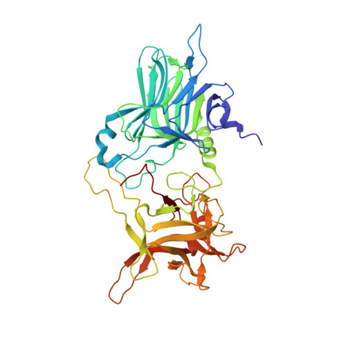
    1Z0H

  • PubMed Abstract: 

    Botulinum neurotoxins comprise seven distinct serotypes (A-G) produced by Clostridium botulinum. The crystal structure of the binding domain of the botulinum neurotoxin type B (BBHc) has been determined to 2A resolution. The overall structure of BBHc is well ordered and similar to that of the binding domain of the holotoxin. However, significant structural changes occur at what would be the interface of translocation and binding domains of the holotoxin. The loop 911-924 shows a maximum displacement of 14.8A at the farthest point. The N-terminal helix reorients and moves by 19.5A from its original position. BBHc is compared with the binding domain of the holotoxin of botulinum type A and B, and the tetanus C-fragment to characterize the heavy chain-carbohydrate interactions. The probable reasons for different binding affinity of botulinum and tetanus toxins are discussed.


  • Organizational Affiliation

    Biology Department, Brookhaven National Laboratory, Upton, NY 11973, USA.


Macromolecules
Find similar proteins by:  (by identity cutoff)  |  3D Structure
Entity ID: 1
MoleculeChains Sequence LengthOrganismDetailsImage
Botulinum neurotoxin type B
A, B
438Clostridium botulinumMutation(s): 0 
Gene Names: botB
EC: 3.4.24.69
UniProt
Find proteins for P10844 (Clostridium botulinum)
Explore P10844 
Go to UniProtKB:  P10844
Entity Groups  
Sequence Clusters30% Identity50% Identity70% Identity90% Identity95% Identity100% Identity
UniProt GroupP10844
Sequence Annotations
Expand
  • Reference Sequence
Experimental Data & Validation

Experimental Data

  • Method: X-RAY DIFFRACTION
  • Resolution: 2.00 Å
  • R-Value Free: 0.282 
  • R-Value Work: 0.234 
  • R-Value Observed: 0.234 
  • Space Group: P 1 21 1
Unit Cell:
Length ( Å )Angle ( ˚ )
a = 68.664α = 90
b = 78.806β = 103.03
c = 87.722γ = 90
Software Package:
Software NamePurpose
CNSrefinement
CBASSdata collection
HKL-2000data scaling
AMoREphasing

Structure Validation

View Full Validation Report


Entry History 

Deposition Data

Revision History  (Full details and data files)

  • Version 1.0: 2005-03-15
    Type: Initial release
  • Version 1.1: 2008-04-30
    Changes: Version format compliance
  • Version 1.2: 2011-07-13
    Changes: Version format compliance
  • Version 1.3: 2023-08-23
    Changes: Data collection, Database references, Refinement description