1YWF

Crystal Structure of Mycobacterium Tuberculosis Protein Tyrosine Phosphatase PtpB


Experimental Data Snapshot

  • Method: X-RAY DIFFRACTION
  • Resolution: 1.71 Å
  • R-Value Free: 0.214 
  • R-Value Work: 0.176 
  • R-Value Observed: 0.178 

wwPDB Validation   3D Report Full Report


This is version 1.3 of the entry. See complete history


Literature

Mycobacterium tuberculosis Protein Tyrosine Phosphatase PtpB Structure Reveals a Diverged Fold and a Buried Active Site.

Grundner, C.Ng, H.L.Alber, T.

(2005) Structure 13: 1625-1634

  • DOI: https://doi.org/10.1016/j.str.2005.07.017
  • Primary Citation of Related Structures:  
    1YWF

  • PubMed Abstract: 

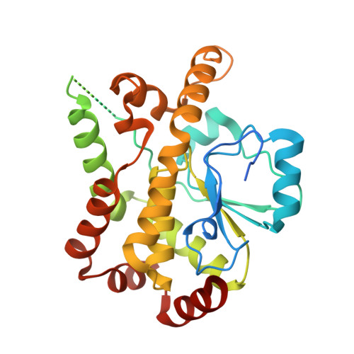
    Intracellular pathogenic bacteria manipulate host signal transduction pathways to facilitate infection. Mycobacterium tuberculosis protein tyrosine phosphatases (PTPs) PtpA and PtpB are thought to be secreted into host cells and interfere with unidentified signals. To illuminate the mechanisms of regulation and substrate recognition, we determined the 1.7 A resolution crystal structure of PtpB in complex with the product phosphate. The protein adopts a simplified PTP fold, which combines features of the conventional PTPs and dual-specificity phosphatases. PtpB shows two unusual elaborations--a disordered, acidic loop and a flexible, two-helix lid that covers the active site--that are specific to mycobacterial orthologs. Biochemical studies suggest that substrate mimicry in the lid may protect the phosphatase from oxidative inactivation. The insertion and deletion of large structural elements in PtpB suggest that, outside the active site module, the PTP family is under unusual selective pressure that promotes changes in overall structure.


  • Organizational Affiliation

    Department of Molecular and Cell Biology, University of California, Berkeley, Berkeley, California 94720, USA.


Macromolecules
Find similar proteins by:  (by identity cutoff)  |  3D Structure
Entity ID: 1
MoleculeChains Sequence LengthOrganismDetailsImage
PHOSPHOTYROSINE PROTEIN PHOSPHATASE PTPB296Mycobacterium tuberculosisMutation(s): 0 
UniProt
Find proteins for P96830 (Mycobacterium tuberculosis (strain CDC 1551 / Oshkosh))
Explore P96830 
Go to UniProtKB:  P96830
Entity Groups  
Sequence Clusters30% Identity50% Identity70% Identity90% Identity95% Identity100% Identity
UniProt GroupP96830
Sequence Annotations
Expand
  • Reference Sequence
Small Molecules
Ligands 1 Unique
IDChains Name / Formula / InChI Key2D Diagram3D Interactions
PO4
Query on PO4

Download Ideal Coordinates CCD File 
B [auth A]PHOSPHATE ION
O4 P
NBIIXXVUZAFLBC-UHFFFAOYSA-K
Experimental Data & Validation

Experimental Data

  • Method: X-RAY DIFFRACTION
  • Resolution: 1.71 Å
  • R-Value Free: 0.214 
  • R-Value Work: 0.176 
  • R-Value Observed: 0.178 
  • Space Group: I 41
Unit Cell:
Length ( Å )Angle ( ˚ )
a = 113.068α = 90
b = 113.068β = 90
c = 53.292γ = 90
Software Package:
Software NamePurpose
REFMACrefinement
HKL-2000data reduction
SCALEPACKdata scaling
SOLVEphasing

Structure Validation

View Full Validation Report


Entry History 

Deposition Data

Revision History  (Full details and data files)

  • Version 1.0: 2005-11-22
    Type: Initial release
  • Version 1.1: 2008-04-30
    Changes: Version format compliance
  • Version 1.2: 2011-07-13
    Changes: Version format compliance
  • Version 1.3: 2024-02-14
    Changes: Data collection, Database references, Derived calculations