1YTM

Crystal structure of phosphoenolpyruvate carboxykinase of Anaerobiospirillum succiniciproducens complexed with ATP, oxalate, magnesium and manganese ions


Experimental Data Snapshot

  • Method: X-RAY DIFFRACTION
  • Resolution: 2.20 Å
  • R-Value Free: 0.251 
  • R-Value Work: 0.215 
  • R-Value Observed: 0.221 

wwPDB Validation   3D Report Full Report


Ligand Structure Quality Assessment 


This is version 1.4 of the entry. See complete history


Literature

Crystal structure of Anaerobiospirillum succiniciproducens PEP carboxykinase reveals an important active site loop

Cotelesage, J.J.H.Prasad, L.Zeikus, J.G.Laivenieks, M.Delbaere, L.T.J.

(2005) Int J Biochem Cell Biol 37: 1829-1837

  • DOI: https://doi.org/10.1016/j.biocel.2005.03.008
  • Primary Citation of Related Structures:  
    1YTM, 1YVY

  • PubMed Abstract: 

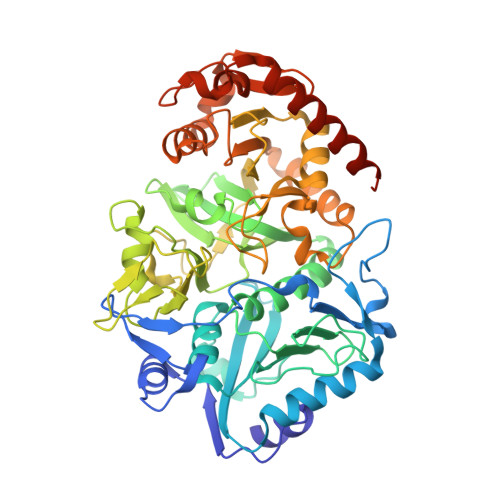
    The 2.2 Angstroms resolution crystal structure of the enzyme phosphoenolpyruvate carboxykinase (PCK) from the bacterium Anaerobiospirillum succiniciproducens complexed with ATP, Mg(2+), Mn(2+) and the transition state analogue oxalate has been solved. The 2.4 Angstroms resolution native structure of A. succiniciproducens PCK has also been determined. It has been found that upon binding of substrate, PCK undergoes a conformational change. Two domains of the molecule fold towards each other, with the substrates and metal ions held in a cleft formed between the two domains. This domain movement is believed to accelerate the reaction PCK catalyzes by forcing bulk solvent molecules out of the active site. Although the crystal structure of A. succiniciproducens PCK with bound substrate and metal ions is related to the structures of PCK from Escherichia coli and Trypanosoma cruzi, it is the first crystal structure from this class of enzymes that clearly shows an important surface loop (residues 383-397) from the C-terminal domain, hydrogen bonding with the peptide backbone of the active site residue Arg60. The interaction between the surface loop and the active site backbone, which is a parallel beta-sheet, seems to be a feature unique of A. succiniciproducens PCK. The association between the loop and the active site is the third type of interaction found in PCK that is thought to play a part in the domain closure. This loop also appears to help accelerate catalysis by functioning as a 'lid' that shields water molecules from the active site.


  • Organizational Affiliation

    Department of Biochemistry, University of Saskatchewan, Saskatoon, Canada.


Macromolecules
Find similar proteins by:  (by identity cutoff)  |  3D Structure
Entity ID: 1
MoleculeChains Sequence LengthOrganismDetailsImage
phosphoenolpyruvate carboxykinase [ATP]
A, B
532Anaerobiospirillum succiniciproducensMutation(s): 0 
EC: 4.1.1.49
UniProt
Find proteins for O09460 (Anaerobiospirillum succiniciproducens)
Explore O09460 
Go to UniProtKB:  O09460
Entity Groups  
Sequence Clusters30% Identity50% Identity70% Identity90% Identity95% Identity100% Identity
UniProt GroupO09460
Sequence Annotations
Expand
  • Reference Sequence
Small Molecules
Ligands 4 Unique
IDChains Name / Formula / InChI Key2D Diagram3D Interactions
ATP
Query on ATP

Download Ideal Coordinates CCD File 
E [auth A],
I [auth B]
ADENOSINE-5'-TRIPHOSPHATE
C10 H16 N5 O13 P3
ZKHQWZAMYRWXGA-KQYNXXCUSA-N
OXD
Query on OXD

Download Ideal Coordinates CCD File 
F [auth A],
J [auth B]
OXALIC ACID
C2 H2 O4
MUBZPKHOEPUJKR-UHFFFAOYSA-N
MN
Query on MN

Download Ideal Coordinates CCD File 
D [auth A],
H [auth B]
MANGANESE (II) ION
Mn
WAEMQWOKJMHJLA-UHFFFAOYSA-N
MG
Query on MG

Download Ideal Coordinates CCD File 
C [auth A],
G [auth B]
MAGNESIUM ION
Mg
JLVVSXFLKOJNIY-UHFFFAOYSA-N
Experimental Data & Validation

Experimental Data

  • Method: X-RAY DIFFRACTION
  • Resolution: 2.20 Å
  • R-Value Free: 0.251 
  • R-Value Work: 0.215 
  • R-Value Observed: 0.221 
  • Space Group: P 21 21 2
Unit Cell:
Length ( Å )Angle ( ˚ )
a = 194.871α = 90
b = 123.219β = 90
c = 48.463γ = 90
Software Package:
Software NamePurpose
HKL-2000data collection
SCALEPACKdata scaling
AMoREphasing
CNSrefinement
HKL-2000data reduction

Structure Validation

View Full Validation Report


Ligand Structure Quality Assessment 


Entry History 

Deposition Data

Revision History  (Full details and data files)

  • Version 1.0: 2006-01-24
    Type: Initial release
  • Version 1.1: 2008-04-30
    Changes: Version format compliance
  • Version 1.2: 2011-07-13
    Changes: Version format compliance
  • Version 1.3: 2021-07-28
    Changes: Derived calculations, Refinement description
  • Version 1.4: 2023-08-23
    Changes: Data collection, Database references, Refinement description