1YT0

Crystal Structure of the Unliganded Form of GRP94, the ER Hsp90: Basis for Nucleotide-Induced Conformational Change, GRP94N(DELTA)41 APO CRYSTAL SOAKED WITH ADP


Experimental Data Snapshot

  • Method: X-RAY DIFFRACTION
  • Resolution: 2.40 Å
  • R-Value Free: 0.264 
  • R-Value Work: 0.228 
  • R-Value Observed: 0.228 

wwPDB Validation   3D Report Full Report


Ligand Structure Quality Assessment 


This is version 1.4 of the entry. See complete history


Literature

Structure of Unliganded GRP94, the Endoplasmic Reticulum Hsp90: BASIS FOR NUCLEOTIDE-INDUCED CONFORMATIONAL CHANGE

Dollins, D.E.Immormino, R.M.Gewirth, D.T.

(2005) J Biol Chem 280: 30438-30447

  • DOI: https://doi.org/10.1074/jbc.M503761200
  • Primary Citation of Related Structures:  
    1YSZ, 1YT0, 1YT1, 1YT2

  • PubMed Abstract: 

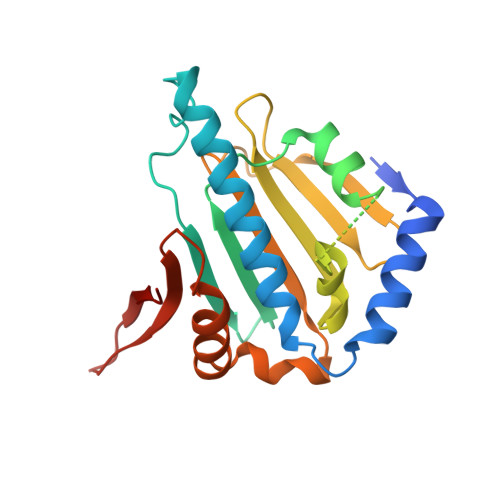
    GRP94, the endoplasmic reticulum paralog of Hsp90, is regulated by adenosine nucleotides that bind to its N-terminal regulatory domain. Because of its weak affinity for nucleotides, the functionally relevant transition in GRP94 is likely to be between the unliganded and nucleotide-bound states. We have determined the structure of the unliganded GRP94 N-domain. The helix 1-4-5 subdomain of the unliganded protein adopts the closed conformation seen in the structure of the protein in complex with inhibitors. This conformation is distinct from the open conformation of the subdomain seen when the protein is bound to ATP or ADP. ADP soaked into crystals of the unliganded protein reveals an intermediate conformation midway between the open and closed states and demonstrates that in GRP94 the conversion between the open and closed states is driven by ligand binding. The direction of the observed movement in GRP94 shows that nucleotides act to open the subdomain elements rather than close them, which is contrary to the motion proposed for Hsp90. These observations support a model where ATP binding dictates the conformation of the N-domain and regulates its ability to form quaternary structural interactions.


  • Organizational Affiliation

    Department of Biochemistry, Duke University Medical Center, Durham, North Carolina 27710, USA.


Macromolecules
Find similar proteins by:  (by identity cutoff)  |  3D Structure
Entity ID: 1
MoleculeChains Sequence LengthOrganismDetailsImage
Endoplasmin236Canis lupus familiarisMutation(s): 0 
Gene Names: TRA1
UniProt
Find proteins for P41148 (Canis lupus familiaris)
Explore P41148 
Go to UniProtKB:  P41148
Entity Groups  
Sequence Clusters30% Identity50% Identity70% Identity90% Identity95% Identity100% Identity
UniProt GroupP41148
Sequence Annotations
Expand
  • Reference Sequence
Small Molecules
Ligands 3 Unique
IDChains Name / Formula / InChI Key2D Diagram3D Interactions
ADP
Query on ADP

Download Ideal Coordinates CCD File 
C [auth A]ADENOSINE-5'-DIPHOSPHATE
C10 H15 N5 O10 P2
XTWYTFMLZFPYCI-KQYNXXCUSA-N
PG4
Query on PG4

Download Ideal Coordinates CCD File 
D [auth A]
E [auth A]
F [auth A]
G [auth A]
H [auth A]
D [auth A],
E [auth A],
F [auth A],
G [auth A],
H [auth A],
I [auth A]
TETRAETHYLENE GLYCOL
C8 H18 O5
UWHCKJMYHZGTIT-UHFFFAOYSA-N
MG
Query on MG

Download Ideal Coordinates CCD File 
B [auth A]MAGNESIUM ION
Mg
JLVVSXFLKOJNIY-UHFFFAOYSA-N
Binding Affinity Annotations 
IDSourceBinding Affinity
ADP BindingDB:  1YT0 IC50: 1.14e+4 (nM) from 1 assay(s)
EC50: 2.10e+4 (nM) from 1 assay(s)
Experimental Data & Validation

Experimental Data

  • Method: X-RAY DIFFRACTION
  • Resolution: 2.40 Å
  • R-Value Free: 0.264 
  • R-Value Work: 0.228 
  • R-Value Observed: 0.228 
  • Space Group: P 21 21 2
Unit Cell:
Length ( Å )Angle ( ˚ )
a = 65.91α = 90
b = 94.86β = 90
c = 42.91γ = 90
Software Package:
Software NamePurpose
CNSrefinement
HKL-2000data reduction
SCALEPACKdata scaling
CNSphasing

Structure Validation

View Full Validation Report


Ligand Structure Quality Assessment 


Entry History 

Deposition Data

Revision History  (Full details and data files)

  • Version 1.0: 2005-06-14
    Type: Initial release
  • Version 1.1: 2008-04-30
    Changes: Version format compliance
  • Version 1.2: 2011-07-13
    Changes: Version format compliance
  • Version 1.3: 2017-08-02
    Changes: Source and taxonomy
  • Version 1.4: 2024-02-14
    Changes: Data collection, Database references, Derived calculations