1YRY

Crystal structure of human PNP complexed with MESG


Experimental Data Snapshot

  • Method: X-RAY DIFFRACTION
  • Resolution: 2.80 Å
  • R-Value Free: 0.290 
  • R-Value Work: 0.207 
  • R-Value Observed: 0.207 

wwPDB Validation   3D Report Full Report


Ligand Structure Quality Assessment 


This is version 1.3 of the entry. See complete history


Literature

Kinetics and crystal structure of human purine nucleoside phosphorylase in complex with 7-methyl-6-thio-guanosine

Silva, R.G.Pereira, J.H.Canduri, F.de Azevedo Jr., W.F.Basso, L.A.Santos, D.S.

(2005) Arch Biochem Biophys 442: 49-58

  • DOI: https://doi.org/10.1016/j.abb.2005.07.021
  • Primary Citation of Related Structures:  
    1YRY

  • PubMed Abstract: 

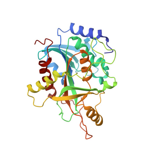
    Purine nucleoside phosphorylase (PNP) catalyzes the reversible phosphorolysis of nucleosides and deoxynucleosides, generating ribose 1-phosphate and the purine base, which is an important step of purine catabolism pathway. The lack of such an activity in humans, owing to a genetic disorder, causes T-cell impairment, and drugs that inhibit this enzyme may have the potential of being utilized as modulators of the immunological system to treat leukemia, autoimmune diseases, and rejection in organ transplantation. Here, we describe kinetics and crystal structure of human PNP in complex with 7-methyl-6-thio-guanosine, a synthetic substrate, which is largely used in activity assays. Analysis of the structure identifies different protein conformational changes upon ligand binding, and comparison of kinetic and structural data permits an understanding of the effects of atomic substitution on key positions of the synthetic substrate and their consequences to enzyme binding and catalysis. Such knowledge may be helpful in designing new PNP inhibitors.


  • Organizational Affiliation

    Centro de Pesquisas em Biologia Molecular e Funcional, Instituto de Pesquisas Biomédicas, PUCRS, Porto Alegre, RS, Brazil.


Macromolecules
Find similar proteins by:  (by identity cutoff)  |  3D Structure
Entity ID: 1
MoleculeChains Sequence LengthOrganismDetailsImage
Purine nucleoside phosphorylaseA [auth E]289Homo sapiensMutation(s): 0 
EC: 2.4.2.1
UniProt & NIH Common Fund Data Resources
Find proteins for P00491 (Homo sapiens)
Explore P00491 
Go to UniProtKB:  P00491
PHAROS:  P00491
GTEx:  ENSG00000198805 
Entity Groups  
Sequence Clusters30% Identity50% Identity70% Identity90% Identity95% Identity100% Identity
UniProt GroupP00491
Sequence Annotations
Expand
  • Reference Sequence
Experimental Data & Validation

Experimental Data

  • Method: X-RAY DIFFRACTION
  • Resolution: 2.80 Å
  • R-Value Free: 0.290 
  • R-Value Work: 0.207 
  • R-Value Observed: 0.207 
  • Space Group: H 3 2
Unit Cell:
Length ( Å )Angle ( ˚ )
a = 138.99α = 90
b = 138.99β = 90
c = 159.85γ = 120
Software Package:
Software NamePurpose
MOSFLMdata reduction
SCALAdata scaling
AMoREphasing
X-PLORrefinement
CCP4data scaling

Structure Validation

View Full Validation Report


Ligand Structure Quality Assessment 


Entry History 

Deposition Data

Revision History  (Full details and data files)

  • Version 1.0: 2006-01-17
    Type: Initial release
  • Version 1.1: 2008-04-30
    Changes: Version format compliance
  • Version 1.2: 2011-07-13
    Changes: Derived calculations, Version format compliance
  • Version 1.3: 2023-10-25
    Changes: Data collection, Database references, Derived calculations, Refinement description