1YP1

Crystal structure of a non-hemorrhagic fibrin(ogen)olytic metalloproteinase from venom of Agkistrodon acutus


Experimental Data Snapshot

  • Method: X-RAY DIFFRACTION
  • Resolution: 1.90 Å
  • R-Value Free: 0.234 
  • R-Value Work: 0.217 
  • R-Value Observed: 0.219 

wwPDB Validation   3D Report Full Report


This is version 1.2 of the entry. See complete history


Literature

Crystal structure of a non-hemorrhagic fibrin(ogen)olytic metalloproteinase complexed with a novel natural tri-peptide inhibitor from venom of Agkistrodon acutus

Lou, Z.Hou, J.Liang, X.Chen, J.Qiu, P.Liu, Y.Li, M.Rao, Z.Yan, G.

(2005) J Struct Biol 152: 195-203

  • DOI: https://doi.org/10.1016/j.jsb.2005.09.006
  • Primary Citation of Related Structures:  
    1YP1

  • PubMed Abstract: 

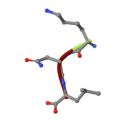
    Thrombotic occlusive diseases pose a great threat to human health. Thrombolytic agents are in widespread use for the dissolution of arterial and venous pathologic thrombi in these kinds of diseases. Snake venom metalloproteinases (SVMPs) can act directly on fibrin/fibrinogen and are therefore potential candidates for therapeutic use against thrombotic occlusive diseases. In this study, we have determined the crystal structure of FII, a novel non-hemorrhagic SVMP isolated from Anhui Agkistrodon acutus snake venom by molecular replacement. The structure reveals that FII is a member of the P-I class SVMPs. The Zn2+ ion essential for hydrolytic activity is found in the active site and is tetrahedrally co-ordinated by three histidine residues and water molecule. Unambiguous electron density for a tri-peptide with sequence KNL is also found located near the active site. Biochemical evidences show that the tri-peptide KNL can inhibit the enzymatic activity of FII.


  • Organizational Affiliation

    Laboratory of Structural Biology, Tsinghua University, Beijing 100084, People's Republic of China.


Macromolecules
Find similar proteins by:  (by identity cutoff)  |  3D Structure
Entity ID: 1
MoleculeChains Sequence LengthOrganismDetailsImage
FII202Deinagkistrodon acutusMutation(s): 0 
EC: 3.4.24
Entity Groups  
Sequence Clusters30% Identity50% Identity70% Identity90% Identity95% Identity100% Identity
Sequence Annotations
Expand
  • Reference Sequence

Find similar proteins by:  Sequence   |   3D Structure  

Entity ID: 2
MoleculeChains Sequence LengthOrganismDetailsImage
KNL3N/AMutation(s): 0 
Sequence Annotations
Expand
  • Reference Sequence
Small Molecules
Ligands 1 Unique
IDChains Name / Formula / InChI Key2D Diagram3D Interactions
ZN
Query on ZN

Download Ideal Coordinates CCD File 
C [auth A]ZINC ION
Zn
PTFCDOFLOPIGGS-UHFFFAOYSA-N
Experimental Data & Validation

Experimental Data

  • Method: X-RAY DIFFRACTION
  • Resolution: 1.90 Å
  • R-Value Free: 0.234 
  • R-Value Work: 0.217 
  • R-Value Observed: 0.219 
  • Space Group: P 31 2 1
Unit Cell:
Length ( Å )Angle ( ˚ )
a = 80.565α = 90
b = 80.565β = 90
c = 66.769γ = 120
Software Package:
Software NamePurpose
HKL-2000data collection
SCALEPACKdata scaling
CNSrefinement
HKL-2000data reduction
CNSphasing

Structure Validation

View Full Validation Report


Entry History 

Deposition Data

Revision History  (Full details and data files)

  • Version 1.0: 2006-01-17
    Type: Initial release
  • Version 1.1: 2008-04-30
    Changes: Version format compliance
  • Version 1.2: 2011-07-13
    Changes: Version format compliance