1YON

Escherichia coli ketopantoate reductase in complex with 2-monophosphoadenosine-5'-diphosphate


Experimental Data Snapshot

  • Method: X-RAY DIFFRACTION
  • Resolution: 1.95 Å
  • R-Value Free: 0.210 
  • R-Value Work: 0.166 
  • R-Value Observed: 0.168 

wwPDB Validation   3D Report Full Report


Ligand Structure Quality Assessment 


This is version 1.4 of the entry. See complete history


Literature

pH-tuneable binding of 2'-phospho-ADP-ribose to ketopantoate reductase: a structural and calorimetric study.

Ciulli, A.Lobley, C.M.Tuck, K.L.Smith, A.G.Blundell, T.L.Abell, C.

(2007) Acta Crystallogr D Biol Crystallogr 63: 171-178

  • DOI: https://doi.org/10.1107/S0907444906044465
  • Primary Citation of Related Structures:  
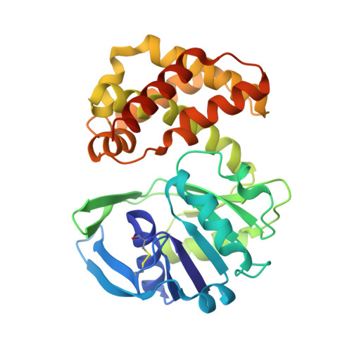
    1YON

  • PubMed Abstract: 

    The crystal structure of Escherichia coli ketopantoate reductase in complex with 2'-monophosphoadenosine 5'-diphosphoribose, a fragment of NADP+ that lacks the nicotinamide ring, is reported. The ligand is bound at the enzyme active site in the opposite orientation to that observed for NADP+, with the adenine ring occupying the lipophilic nicotinamide pocket. Isothermal titration calorimetry with R31A and N98A mutants of the enzyme is used to show that the unusual ;reversed binding mode' observed in the crystal is triggered by changes in the protonation of binding groups at low pH. This research has important implications for fragment-based approaches to drug design, namely that the crystallization conditions and the chemical modification of ligands can have unexpected effects on the binding modes.


  • Organizational Affiliation

    University Chemical Laboratory, Lensfield Road, Cambridge CB2 1EW, England.


Macromolecules
Find similar proteins by:  (by identity cutoff)  |  3D Structure
Entity ID: 1
MoleculeChains Sequence LengthOrganismDetailsImage
2-dehydropantoate 2-reductase303Escherichia coliMutation(s): 0 
Gene Names: panE
EC: 1.1.1.169
UniProt
Find proteins for P0A9J4 (Escherichia coli (strain K12))
Explore P0A9J4 
Go to UniProtKB:  P0A9J4
Entity Groups  
Sequence Clusters30% Identity50% Identity70% Identity90% Identity95% Identity100% Identity
UniProt GroupP0A9J4
Sequence Annotations
Expand
  • Reference Sequence
Small Molecules
Ligands 1 Unique
IDChains Name / Formula / InChI Key2D Diagram3D Interactions
A2R
Query on A2R

Download Ideal Coordinates CCD File 
B [auth A][(2R,3R,4R,5R)-5-(6-AMINO-9H-PURIN-9-YL)-3-HYDROXY-4-(PHOSPHONOOXY)TETRAHYDROFURAN-2-YL]METHYL [(2R,3S,4R,5R)-3,4,5-TRIHYDROXYTETRAHYDROFURAN-2-YL]METHYL DIHYDROGEN DIPHOSPHATE
C15 H24 N5 O17 P3
ICNHOLCERMYLRZ-KEOHHSTQSA-N
Binding Affinity Annotations 
IDSourceBinding Affinity
A2R Binding MOAD:  1YON Kd: 6.10e+4 (nM) from 1 assay(s)
APX PDBBind:  1YON Kd: 6.10e+4 (nM) from 1 assay(s)
Experimental Data & Validation

Experimental Data

  • Method: X-RAY DIFFRACTION
  • Resolution: 1.95 Å
  • R-Value Free: 0.210 
  • R-Value Work: 0.166 
  • R-Value Observed: 0.168 
  • Space Group: P 21 21 21
Unit Cell:
Length ( Å )Angle ( ˚ )
a = 60.318α = 90
b = 65.833β = 90
c = 98.207γ = 90
Software Package:
Software NamePurpose
REFMACrefinement
DENZOdata reduction
SCALEPACKdata scaling
AMoREphasing

Structure Validation

View Full Validation Report


Ligand Structure Quality Assessment 


Entry History 

Deposition Data

Revision History  (Full details and data files)

  • Version 1.0: 2006-04-18
    Type: Initial release
  • Version 1.1: 2008-04-30
    Changes: Version format compliance
  • Version 1.2: 2011-07-13
    Changes: Version format compliance
  • Version 1.3: 2014-11-19
    Changes: Non-polymer description
  • Version 1.4: 2023-10-25
    Changes: Data collection, Database references, Derived calculations, Refinement description