1YNY

Molecular Structure of D-Hydantoinase from a Bacillus sp. AR9: Evidence for mercury inhibition


Experimental Data Snapshot

  • Method: X-RAY DIFFRACTION
  • Resolution: 2.30 Å
  • R-Value Free: 0.243 
  • R-Value Work: 0.199 

wwPDB Validation   3D Report Full Report


This is version 1.4 of the entry. See complete history


Literature

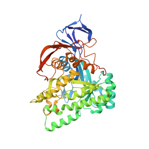
Molecular structure of D-hydantoinase from Bacillus sp. AR9: evidence for mercury inhibition.

Radha Kishan, K.V.Vohra, R.M.Ganesan, K.Agrawal, V.Sharma, V.M.Sharma, R.

(2005) J Mol Biol 347: 95-105

  • DOI: https://doi.org/10.1016/j.jmb.2005.01.025
  • Primary Citation of Related Structures:  
    1YNY

  • PubMed Abstract: 

    Stereospecific conversion of hydantoins into their carbamoyl acid derivatives could be achieved by using the enzyme hydantoinase. Specific hydantoinases convert either the D-form or the L-form of the hydantoin and the amino acids responsible for stereospecificity have not been identified. Structural studies on hydantoinases from a few bacterial species were published recently. The structure of a thermostable D-hydantoinase from Bacillus sp. AR9 (bar9HYD) was solved to 2.3 angstroms resolution. The usual modification of carboxylation of the active-site residue Lys150 did not happen in bar9HYD. Two manganese ions were modelled in the active site. Through biochemical studies, it was shown that mercury inhibits the activity of the enzyme. The mercury derivative provided some information about the binding site of the mercuric inhibitors and a possible reason for inhibition is presented.


  • Organizational Affiliation

    Institute of Microbial Technology, Sector 39-A, Chandigarh 160 036, India. kishan@imtech.res.in


Macromolecules
Find similar proteins by:  (by identity cutoff)  |  3D Structure
Entity ID: 1
MoleculeChains Sequence LengthOrganismDetailsImage
D-Hydantoinase
A, B
461Bacillus sp. AR9Mutation(s): 0 
EC: 3.5.2.2
UniProt
Find proteins for Q5DLU2 (Bacillus sp. AR9)
Explore Q5DLU2 
Go to UniProtKB:  Q5DLU2
Entity Groups  
Sequence Clusters30% Identity50% Identity70% Identity90% Identity95% Identity100% Identity
UniProt GroupQ5DLU2
Sequence Annotations
Expand
  • Reference Sequence
Experimental Data & Validation

Experimental Data

  • Method: X-RAY DIFFRACTION
  • Resolution: 2.30 Å
  • R-Value Free: 0.243 
  • R-Value Work: 0.199 
  • Space Group: P 64
Unit Cell:
Length ( Å )Angle ( ˚ )
a = 129.54α = 90
b = 129.54β = 90
c = 102.85γ = 120
Software Package:
Software NamePurpose
MAR345data collection
SCALEPACKdata scaling
AMoREphasing
CNSrefinement

Structure Validation

View Full Validation Report


Entry History 

Deposition Data

Revision History  (Full details and data files)

  • Version 1.0: 2005-03-01
    Type: Initial release
  • Version 1.1: 2008-04-30
    Changes: Version format compliance
  • Version 1.2: 2011-07-13
    Changes: Source and taxonomy, Version format compliance
  • Version 1.3: 2017-10-11
    Changes: Refinement description
  • Version 1.4: 2023-08-23
    Changes: Data collection, Database references, Derived calculations, Refinement description