1YL7

the crystal structure of Mycobacterium tuberculosis dihydrodipicolinate reductase (Rv2773c) in complex with NADH (crystal form C)


Experimental Data Snapshot

  • Method: X-RAY DIFFRACTION
  • Resolution: 2.34 Å
  • R-Value Free: 0.238 
  • R-Value Work: 0.181 
  • R-Value Observed: 0.183 

wwPDB Validation   3D Report Full Report


Ligand Structure Quality Assessment 


This is version 1.5 of the entry. See complete history


Literature

The structure of dihydrodipicolinate reductase (DapB) from Mycobacterium tuberculosis in three crystal forms.

Janowski, R.Kefala, G.Weiss, M.S.

(2010) Acta Crystallogr D Biol Crystallogr 66: 61-72

  • DOI: https://doi.org/10.1107/S0907444909043960
  • Primary Citation of Related Structures:  
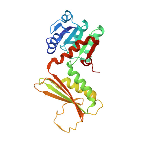
    1YL5, 1YL6, 1YL7

  • PubMed Abstract: 

    Dihydrodipicolinate reductase (DHDPR, DapB) is an enzyme that belongs to the L-lysine biosynthetic pathway. DHDPR reduces the alpha,beta-unsaturated cyclic imine 2,3-dihydrodipicolinic acid to yield the compound 2,3,4,5-tetrahydrodipicolinic acid in a pyridine nucleotide-dependent reaction. The substrate of this reaction is the unstable product of the preceding enzyme dihydrodipicolinate synthase (DHDPS, DapA). Here, the structure of apo-DHDPR from Mycobacterium tuberculosis is reported in two orthorhombic crystal forms, as well as the structure of DHDPR from M. tuberculosis in complex with NADH in a monoclinic crystal form. A comparison of the results with previously solved structures of this enzyme shows that DHDPR undergoes a major conformational change upon binding of its cofactor. This conformational change can be interpreted as one of the low-frequency normal modes of the structure.


  • Organizational Affiliation

    EMBL Hamburg Outstation, c/o DESY, Notkestrasse 85, D-22603 Hamburg, Germany.


Macromolecules
Find similar proteins by:  (by identity cutoff)  |  3D Structure
Entity ID: 1
MoleculeChains Sequence LengthOrganismDetailsImage
Dihydrodipicolinate reductase
A, B, C, D, E
A, B, C, D, E, F, G, H
245Mycobacterium tuberculosisMutation(s): 0 
Gene Names: dapB
EC: 1.3.1.26
UniProt
Find proteins for P9WP23 (Mycobacterium tuberculosis (strain ATCC 25618 / H37Rv))
Explore P9WP23 
Go to UniProtKB:  P9WP23
Entity Groups  
Sequence Clusters30% Identity50% Identity70% Identity90% Identity95% Identity100% Identity
UniProt GroupP9WP23
Sequence Annotations
Expand
  • Reference Sequence
Small Molecules
Ligands 2 Unique
IDChains Name / Formula / InChI Key2D Diagram3D Interactions
NAI
Query on NAI

Download Ideal Coordinates CCD File 
J [auth A]
L [auth B]
M [auth C]
O [auth D]
Q [auth E]
J [auth A],
L [auth B],
M [auth C],
O [auth D],
Q [auth E],
S [auth F],
U [auth G],
V [auth H]
1,4-DIHYDRONICOTINAMIDE ADENINE DINUCLEOTIDE
C21 H29 N7 O14 P2
BOPGDPNILDQYTO-NNYOXOHSSA-N
MG
Query on MG

Download Ideal Coordinates CCD File 
I [auth A]
K [auth B]
N [auth D]
P [auth E]
R [auth F]
I [auth A],
K [auth B],
N [auth D],
P [auth E],
R [auth F],
T [auth G]
MAGNESIUM ION
Mg
JLVVSXFLKOJNIY-UHFFFAOYSA-N
Experimental Data & Validation

Experimental Data

  • Method: X-RAY DIFFRACTION
  • Resolution: 2.34 Å
  • R-Value Free: 0.238 
  • R-Value Work: 0.181 
  • R-Value Observed: 0.183 
  • Space Group: C 1 2 1
Unit Cell:
Length ( Å )Angle ( ˚ )
a = 238.6α = 90
b = 67.52β = 117.08
c = 154.49γ = 90
Software Package:
Software NamePurpose
REFMACrefinement
MAR345data collection
SCALEPACKdata scaling
MOLREPphasing

Structure Validation

View Full Validation Report


Ligand Structure Quality Assessment 


Entry History 

Deposition Data

Revision History  (Full details and data files)

  • Version 1.0: 2006-01-17
    Type: Initial release
  • Version 1.1: 2008-04-30
    Changes: Version format compliance
  • Version 1.2: 2011-07-13
    Changes: Advisory, Version format compliance
  • Version 1.3: 2013-10-30
    Changes: Non-polymer description
  • Version 1.4: 2017-10-11
    Changes: Refinement description
  • Version 1.5: 2023-08-23
    Changes: Data collection, Database references, Derived calculations, Refinement description