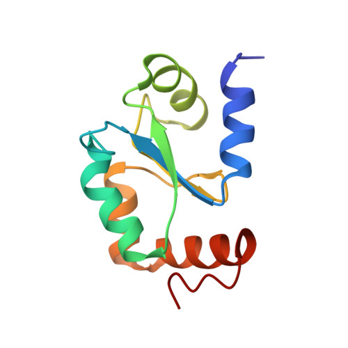
Molecular mapping of functionalities in the solution structure of reduced Grx4, a monothiol glutaredoxin from Escherichia coli.
Fladvad, M., Bellanda, M., Fernandes, A.P., Mammi, S., Vlamis-Gardikas, A., Holmgren, A., Sunnerhagen, M.(2005) J Biol Chem 280: 24553-24561
- PubMed: 15840565
- DOI: https://doi.org/10.1074/jbc.M500679200
- Primary Citation of Related Structures:
1YKA - PubMed Abstract:
The ubiquitous glutaredoxin protein family is present in both prokaryotes and eukaryotes, and is closely related to the thioredoxins, which reduce their substrates using a dithiol mechanism as part of the cellular defense against oxidative stress. Recently identified monothiol glutaredoxins, which must use a different functional mechanism, appear to be essential in both Escherichia coli and yeast and are well conserved in higher order genomes. We have employed high resolution NMR to determine the three-dimensional solution structure of a monothiol glutaredoxin, the reduced E. coli Grx4. The Grx4 structure comprises a glutaredoxin-like alpha-beta fold, founded on a limited set of strictly conserved and structurally critical residues. A tight hydrophobic core, together with a stringent set of secondary structure elements, is thus likely to be present in all monothiol glutaredoxins. A set of exposed and conserved residues form a surface region, implied in glutathione binding from a known structure of E. coli Grx3. The absence of glutaredoxin activity in E. coli Grx4 can be understood based on small but significant differences in the glutathione binding region, and through the lack of a conserved second GSH binding site. MALDI experiments suggest that disulfide formation on glutathionylation is accompanied by significant structural changes, in contrast with dithiol thioredoxins and glutaredoxins, where differences between oxidized and reduced forms are subtle and local. Structural and functional implications are discussed with particular emphasis on identifying common monothiol glutaredoxin properties in substrate specificity and ligand binding events, linking the thioredoxin and glutaredoxin systems.
Organizational Affiliation:
Department of Medical Biochemistry and Biophysics, Karolinska Institutet, S-171 77 Stockholm, Sweden.