A Structural Basis for the Role of Nucleotide Specifying Residues in Regulating the Oligomerization of the Rv1625c Adenylyl Cyclase from M.tuberculosis
Ketkar, A.D., Shenoy, A.R., Ramagopal, U.A., Visweswariah, S.S., Suguna, K.(2006) J Mol Biol 356: 904-916
- PubMed: 16403515
- DOI: https://doi.org/10.1016/j.jmb.2005.12.017
- Primary Citation of Related Structures:
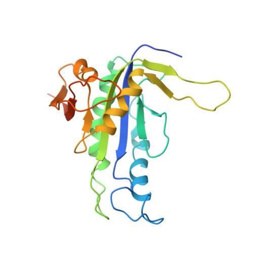
1YK9 - PubMed Abstract:
The Rv1625c Class III adenylyl cyclase from Mycobacterium tuberculosis is a homodimeric enzyme with two catalytic centers at the dimer interface, and shows sequence similarity with the mammalian adenylyl and guanylyl cyclases. Mutation of the substrate-specifying residues in the catalytic domain of Rv1625c, either independently or together, to those present in guanylyl cyclases not only failed to confer guanylyl cyclase activity to the protein, but also severely abrogated the adenylyl cyclase activity of the enzyme. Biochemical analysis revealed alterations in the behavior of the mutants on ion-exchange chromatography, indicating differences in the surface-exposed charge upon mutation of substrate-specifying residues. The mutant proteins showed alterations in oligomeric status as compared to the wild-type enzyme, and differing abilities to heterodimerize with the wild-type protein. The crystal structure of a mutant has been solved to a resolution of 2.7A. On the basis of the structure, and additional biochemical studies, we provide possible reasons for the altered properties of the mutant proteins, as well as highlight unique structural features of the Rv1625c adenylyl cyclase.
Organizational Affiliation:
Department of Molecular Reproduction, Development and Genetics, Indian Institute of Science, Bangalore, 560012, India.