1YIO

Crystallographic structure of response regulator StyR from Pseudomonas fluorescens


Experimental Data Snapshot

  • Method: X-RAY DIFFRACTION
  • Resolution: 2.20 Å
  • R-Value Free: 0.275 
  • R-Value Work: 0.201 
  • R-Value Observed: 0.205 

wwPDB Validation   3D Report Full Report


This is version 1.3 of the entry. See complete history


Literature

An Active-like Structure in the Unphosphorylated StyR Response Regulator Suggests a Phosphorylation- Dependent Allosteric Activation Mechanism

Milani, M.Leoni, L.Rampioni, G.Zennaro, E.Ascenzi, P.Bolognesi, M.

(2005) Structure 13: 1289-1297

  • DOI: https://doi.org/10.1016/j.str.2005.05.014
  • Primary Citation of Related Structures:  
    1YIO, 1ZN2

  • PubMed Abstract: 

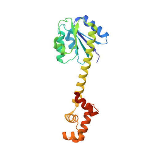
    StyR belongs to the FixJ subfamily of signal transduction response regulators; it controls transcription of the styABCD operon coding for styrene catabolism in Pseudomonas fluorescens ST. The crystal structure of unphosphorylated StyR is reported at 2.2 A resolution. StyR is composed of an N-terminal regulatory domain (StyR-N) and a C-terminal DNA binding domain (StyR-C). The two domains are separated by an elongated linker alpha helix (34 residues), a new feature in known response regulator structures. StyR-C is structured similarly to the DNA binding domain of the response regulator NarL. StyR-N shows structural reorganization of the phosphate receiving region involved in activation/homodimerization: specific residues adopt an "active-like" conformation, and the alpha4 helix, involved in dimerization of the homologous FixJ response regulator, is trimmed to just one helical turn. Overall, structural considerations suggest that phosphorylation may act as an allosteric switch, shifting a preexisting StyR equilibrium toward the active, dimeric, DNA binding form.


  • Organizational Affiliation

    Giannina Gaslini Institute and INFM, Largo G. Gaslini 5, I-16147 Genova, Italy.


Macromolecules
Find similar proteins by:  (by identity cutoff)  |  3D Structure
Entity ID: 1
MoleculeChains Sequence LengthOrganismDetailsImage
response regulatory protein208Pseudomonas fluorescensMutation(s): 0 
UniProt
Find proteins for O30989 (Pseudomonas fluorescens)
Explore O30989 
Go to UniProtKB:  O30989
Entity Groups  
Sequence Clusters30% Identity50% Identity70% Identity90% Identity95% Identity100% Identity
UniProt GroupO30989
Sequence Annotations
Expand
  • Reference Sequence
Experimental Data & Validation

Experimental Data

  • Method: X-RAY DIFFRACTION
  • Resolution: 2.20 Å
  • R-Value Free: 0.275 
  • R-Value Work: 0.201 
  • R-Value Observed: 0.205 
  • Space Group: P 1 21 1
Unit Cell:
Length ( Å )Angle ( ˚ )
a = 41.196α = 90
b = 35.679β = 99.15
c = 73.877γ = 90
Software Package:
Software NamePurpose
REFMACrefinement
DENZOdata reduction
SCALEPACKdata scaling
MOLREPphasing

Structure Validation

View Full Validation Report


Entry History 

Deposition Data

Revision History  (Full details and data files)

  • Version 1.0: 2005-09-27
    Type: Initial release
  • Version 1.1: 2008-04-30
    Changes: Version format compliance
  • Version 1.2: 2011-07-13
    Changes: Version format compliance
  • Version 1.3: 2024-03-13
    Changes: Data collection, Database references, Derived calculations