The catalytic copper of Peptidylglycine alpha-Hydroxylating Monooxygenase also plays a critical structural role.
Siebert, X., Eipper, B.A., Mains, R.E., Prigge, S.T., Blackburn, N.J., Amzel, L.M.(2005) Biophys J 89: 3312-3319
- PubMed: 16100265
- DOI: https://doi.org/10.1529/biophysj.105.066100
- Primary Citation of Related Structures:
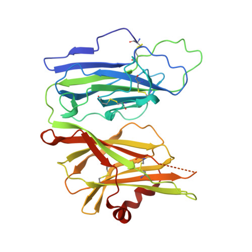
1YI9, 1YIP, 1YJK, 1YJL - PubMed Abstract:
Many bioactive peptides require amidation of their carboxy terminus to exhibit full biological activity. Peptidylglycine alpha-hydroxylating monooxygenase (PHM; EC 1.14.17.3), the enzyme that catalyzes the first of the two steps of this reaction, is composed of two domains, each of which binds one copper atom (CuH and CuM). The CuM site includes Met(314) and two His residues as ligands. Mutation of Met(314) to Ile inactivates PHM, but has only a minimal effect on the EXAFS spectrum of the oxidized enzyme, implying that it contributes only marginally to stabilization of the CuM site. To characterize the role of Met(314) as a CuM ligand, we determined the structure of the Met(314)Ile-PHM mutant. Since the mutant protein failed to crystallize in the conditions of the original wild-type protein, this structure determination required finding a new crystal form. The Met(314)Ile-PHM mutant structure confirms that the mutation does not abolish CuM binding to the enzyme, but causes other structural perturbations that affect the overall stability of the enzyme and the integrity of the CuH site. To eliminate possible effects of crystal contacts, we redetermined the structure of wt-PHM in the Met(314)Ile-PHM crystal form and showed that it does not differ from the structure of wild-type (wt)-PHM in the original crystals. Met(314)Ile-PHM was also shown to be less stable than wt-PHM by differential scanning calorimetry. Both structural and calorimetric studies point to a structural role for the CuM site, in addition to its established catalytic role.
Organizational Affiliation:
Biophysics and Biophysical Chemistry, Johns Hopkins University School of Medicine, Baltimore, MD, USA.