Structure of PEP carboxykinase from the succinate-producing Actinobacillus succinogenes: a new conserved active-site motif.
Leduc, Y.A., Prasad, L., Laivenieks, M., Zeikus, J.G., Delbaere, L.T.(2005) Acta Crystallogr D Biol Crystallogr 61: 903-912
- PubMed: 15983413
- DOI: https://doi.org/10.1107/S0907444905008723
- Primary Citation of Related Structures:
1YGG, 1YLH - PubMed Abstract:
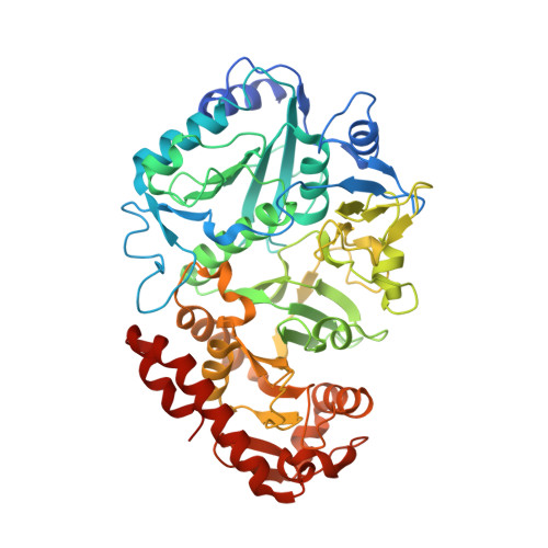
Actinobacillus succinogenes can produce, via fermentation, high concentrations of succinate, an important industrial commodity. A key enzyme in this pathway is phosphoenolpyruvate carboxykinase (PCK), which catalyzes the production of oxaloacetate from phosphoenolpyruvate and carbon dioxide, with the concomitant conversion of adenosine 5'-diphosphate to adenosine 5'-triphosphate. 1.85 and 1.70 A resolution structures of the native and a pyruvate/Mn(2+)/phosphate complex have been solved, respectively. The structure of the complex contains sulfhydryl reducing agents covalently bound to three cysteine residues via disulfide bonds. One of these cysteine residues (Cys285) is located in the active-site cleft and may be analogous to the putative reactive cysteine of PCK from Trypanosoma cruzi. Cys285 is also part of a previously unreported conserved motif comprising residues 280-287 and containing the pattern NXEXGXY(/F)A(/G); this new motif appears to have a structural role in stabilizing and positioning side chains that bind substrates and metal ions. The first few residues of this motif connect the two domains of the enzyme and a fulcrum point appears to be located near Asn280. In addition, an active-site Asp residue forms two coordinate bonds with the Mn(2+) ion present in the structure of the complex in a symmetrical bidentate manner, unlike in other PCK structures that contain a manganese ion.
Organizational Affiliation:
Department of Biochemistry, University of Saskatchewan, Saskatoon, Saskatchewan S7N 5E5, Canada.