1YGC

Short Factor VIIa with a small molecule inhibitor


Experimental Data Snapshot

  • Method: X-RAY DIFFRACTION
  • Resolution: 2.00 Å
  • R-Value Free: 0.205 
  • R-Value Work: 0.188 
  • R-Value Observed: 0.189 

wwPDB Validation   3D Report Full Report


Ligand Structure Quality Assessment 


This is version 1.2 of the entry. See complete history


Literature

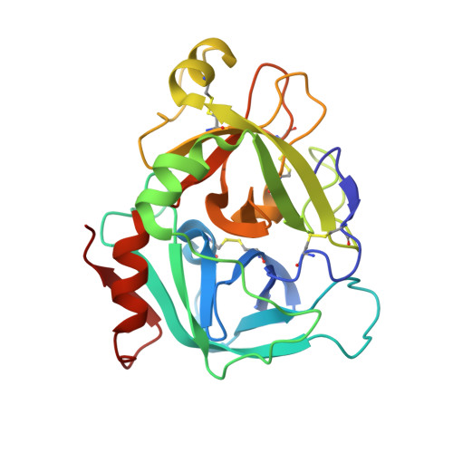
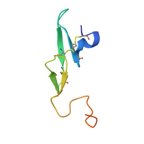
A selective, slow binding inhibitor of factor VIIa binds to a nonstandard active site conformation and attenuates thrombus formation in vivo.

Olivero, A.G.Eigenbrot, C.Goldsmith, R.Robarge, K.Artis, D.R.Flygare, J.Rawson, T.Sutherlin, D.P.Kadkhodayan, S.Beresini, M.Elliott, L.O.DeGuzman, G.G.Banner, D.W.Ultsch, M.Marzec, U.Hanson, S.R.Refino, C.Bunting, S.Kirchhofer, D.

(2005) J Biol Chem 280: 9160-9169

  • DOI: https://doi.org/10.1074/jbc.M409068200
  • Primary Citation of Related Structures:  
    1YGC

  • PubMed Abstract: 

    The serine protease factor VIIa (FVIIa) in complex with its cellular cofactor tissue factor (TF) initiates the blood coagulation reactions. TF.FVIIa is also implicated in thrombosis-related disorders and constitutes an appealing therapeutic target for treatment of cardiovascular diseases. To this end, we generated the FVIIa active site inhibitor G17905, which displayed great potency toward TF.FVIIa (Ki = 0.35 +/- 0.11 nM). G17905 did not appreciably inhibit 12 of the 14 examined trypsin-like serine proteases, consistent with its TF.FVIIa-specific activity in clotting assays. The crystal structure of the FVIIa.G17905 complex provides insight into the molecular basis of the high selectivity. It shows that, compared with other serine proteases, FVIIa is uniquely equipped to accommodate conformational disturbances in the Gln217-Gly219 region caused by the ortho-hydroxy group of the inhibitor's aminobenzamidine moiety located in the S1 recognition pocket. Moreover, the structure revealed a novel, nonstandard conformation of FVIIa active site in the region of the oxyanion hole, a "flipped" Lys192-Gly193 peptide bond. Macromolecular substrate activation assays demonstrated that G17905 is a noncompetitive, slow-binding inhibitor. Nevertheless, G17905 effectively inhibited thrombus formation in a baboon arterio-venous shunt model, reducing platelet and fibrin deposition by approximately 70% at 0.4 mg/kg + 0.1 mg/kg/min infusion. Therefore, the in vitro potency of G17905, characterized by slow binding kinetics, correlated with efficacious antithrombotic activity in vivo.


  • Organizational Affiliation

    Department of Medicinal Chemistry, Genentech, Inc., South San Francisco, California 94080, USA.


Macromolecules
Find similar proteins by:  (by identity cutoff)  |  3D Structure
Entity ID: 1
MoleculeChains Sequence LengthOrganismDetailsImage
Coagulation factor VIIA [auth H]254Homo sapiensMutation(s): 0 
Gene Names: F7
EC: 3.4.21.21
UniProt & NIH Common Fund Data Resources
Find proteins for P08709 (Homo sapiens)
Explore P08709 
Go to UniProtKB:  P08709
PHAROS:  P08709
GTEx:  ENSG00000057593 
Entity Groups  
Sequence Clusters30% Identity50% Identity70% Identity90% Identity95% Identity100% Identity
UniProt GroupP08709
Sequence Annotations
Expand
  • Reference Sequence
Find similar proteins by:  (by identity cutoff)  |  3D Structure
Entity ID: 2
MoleculeChains Sequence LengthOrganismDetailsImage
Coagulation factor VIIB [auth L]63Homo sapiensMutation(s): 0 
Gene Names: F7
UniProt & NIH Common Fund Data Resources
Find proteins for P08709 (Homo sapiens)
Explore P08709 
Go to UniProtKB:  P08709
PHAROS:  P08709
GTEx:  ENSG00000057593 
Entity Groups  
Sequence Clusters30% Identity50% Identity70% Identity90% Identity95% Identity100% Identity
UniProt GroupP08709
Sequence Annotations
Expand
  • Reference Sequence
Small Molecules
Ligands 3 Unique
IDChains Name / Formula / InChI Key2D Diagram3D Interactions
905
Query on 905

Download Ideal Coordinates CCD File 
K [auth H](R)-4-[2-(3-AMINO-BENZENESULFONYLAMINO)-1-(3,5-DIETHOXY-2-FLUOROPHENYL)-2-OXO-ETHYLAMINO]-2-HYDROXY-BENZAMIDINE
C25 H28 F N5 O6 S
MLPXCKVDBGAVRJ-HSZRJFAPSA-N
SO4
Query on SO4

Download Ideal Coordinates CCD File 
E [auth H]
F [auth H]
G [auth H]
H
I [auth H]
E [auth H],
F [auth H],
G [auth H],
H,
I [auth H],
J [auth H],
L
SULFATE ION
O4 S
QAOWNCQODCNURD-UHFFFAOYSA-L
CA
Query on CA

Download Ideal Coordinates CCD File 
C [auth H],
D [auth H]
CALCIUM ION
Ca
BHPQYMZQTOCNFJ-UHFFFAOYSA-N
Binding Affinity Annotations 
IDSourceBinding Affinity
905 Binding MOAD:  1YGC Ki: 0.35 (nM) from 1 assay(s)
BindingDB:  1YGC Ki: 0.35 (nM) from 1 assay(s)
Experimental Data & Validation

Experimental Data

  • Method: X-RAY DIFFRACTION
  • Resolution: 2.00 Å
  • R-Value Free: 0.205 
  • R-Value Work: 0.188 
  • R-Value Observed: 0.189 
  • Space Group: P 41 21 2
Unit Cell:
Length ( Å )Angle ( ˚ )
a = 95.22α = 90
b = 95.22β = 90
c = 116.32γ = 90
Software Package:
Software NamePurpose
CNXrefinement
DENZOdata reduction
SCALEPACKdata scaling
AMoREphasing

Structure Validation

View Full Validation Report


Ligand Structure Quality Assessment 


Entry History 

Deposition Data

Revision History  (Full details and data files)

  • Version 1.0: 2005-01-18
    Type: Initial release
  • Version 1.1: 2008-04-30
    Changes: Version format compliance
  • Version 1.2: 2011-07-13
    Changes: Version format compliance