1YFO

RECEPTOR PROTEIN TYROSINE PHOSPHATASE ALPHA, DOMAIN 1 FROM MOUSE


Experimental Data Snapshot

  • Method: X-RAY DIFFRACTION
  • Resolution: 2.25 Å
  • R-Value Free: 0.251 
  • R-Value Work: 0.184 
  • R-Value Observed: 0.184 

wwPDB Validation   3D Report Full Report


This is version 1.3 of the entry. See complete history


Literature

Structural basis for inhibition of receptor protein-tyrosine phosphatase-alpha by dimerization.

Bilwes, A.M.den Hertog, J.Hunter, T.Noel, J.P.

(1996) Nature 382: 555-559

  • DOI: https://doi.org/10.1038/382555a0
  • Primary Citation of Related Structures:  
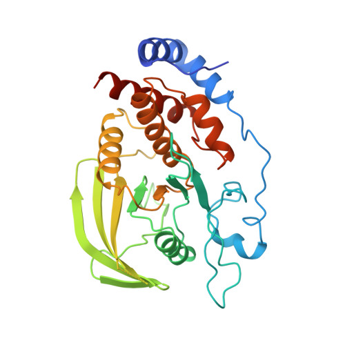
    1YFO

  • PubMed Abstract: 

    Receptor-like protein-tyrosine phosphatases (RPTPs), like their non-receptor counterparts, regulate the level of phosphotyrosine-containing proteins derived from the action of protein-tyrosine kinases. RPTPs are type-I integral membrane proteins which contain one or two catalytic domains in their cytoplasmic region. It is not known whether extracellular ligands regulate the activity of RPTPs. Here we describe the crystal structure of the membrane-proximal catalytic domain (D1) of a typical RPTP, murine RPTP alpha. Significant structural deviations from the PTP1B fold reside within the amino-terminal helix-turn-helix segment of RPTPalphaD1 (residues 214 to 242) and a distinctive two-stranded beta-sheet formed between residues 211-213 and 458-461. The turn of the N-terminal segment inserts into the active site of a dyad-related D1 monomer. On the basis of two independent crystal structures, sequence alignments, and the reported biological activity of EGF receptor/CD45 chimaeras, we propose that dimerization and active-site blockage is a physiologically important mechanism for downregulating the catalytic activity of RPTPalpha and other RPTPs.


  • Organizational Affiliation

    Structural Biology Laboratory, The Salk Institute for Biological Studies, La Jolla, California 92037, USA.


Macromolecules
Find similar proteins by:  (by identity cutoff)  |  3D Structure
Entity ID: 1
MoleculeChains Sequence LengthOrganismDetailsImage
RECEPTOR PROTEIN TYROSINE PHOSPHATASE ALPHA
A, B
302Mus musculusMutation(s): 0 
EC: 3.1.3.48
UniProt & NIH Common Fund Data Resources
Find proteins for P18052 (Mus musculus)
Explore P18052 
Go to UniProtKB:  P18052
IMPC:  MGI:97808
Entity Groups  
Sequence Clusters30% Identity50% Identity70% Identity90% Identity95% Identity100% Identity
UniProt GroupP18052
Sequence Annotations
Expand
  • Reference Sequence
Experimental Data & Validation

Experimental Data

  • Method: X-RAY DIFFRACTION
  • Resolution: 2.25 Å
  • R-Value Free: 0.251 
  • R-Value Work: 0.184 
  • R-Value Observed: 0.184 
  • Space Group: P 1 21 1
Unit Cell:
Length ( Å )Angle ( ˚ )
a = 42.19α = 90
b = 119.84β = 110.08
c = 61.18γ = 90
Software Package:
Software NamePurpose
X-PLORmodel building
X-PLORrefinement
DENZOdata reduction
SCALEPACKdata scaling
X-PLORphasing

Structure Validation

View Full Validation Report


Entry History 

Deposition Data

Revision History  (Full details and data files)

  • Version 1.0: 1997-04-01
    Type: Initial release
  • Version 1.1: 2008-03-24
    Changes: Version format compliance
  • Version 1.2: 2011-07-13
    Changes: Version format compliance
  • Version 1.3: 2023-08-09
    Changes: Database references, Other, Refinement description