1YFE

Crystal structure of apo fumarase C from Escherichia coli


Experimental Data Snapshot

  • Method: X-RAY DIFFRACTION
  • Resolution: 2.19 Å
  • R-Value Free: 0.210 
  • R-Value Work: 0.203 
  • R-Value Observed: 0.203 

wwPDB Validation   3D Report Full Report


This is version 1.4 of the entry. See complete history


Literature

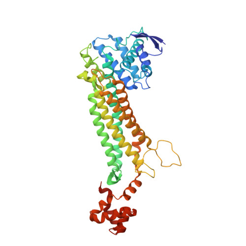
Structure of free fumarase C from Escherichia coli.

Weaver, T.

(2005) Acta Crystallogr D Biol Crystallogr 61: 1395-1401

  • DOI: https://doi.org/10.1107/S0907444905024194
  • Primary Citation of Related Structures:  
    1YFE

  • PubMed Abstract: 

    Previous crystal structures of fumarase C from Escherichia coli have noted two occupied dicarboxylate-binding sites termed the active site and the B site. Here, the first known fumarase C structure is reported in which both sites are unoccupied by bound ligand. This so-called ;free' crystal form shows conservation of the active-site water in a similar orientation to that reported in other fumarase C crystal structures. More importantly, a shift of His129 has been observed at the B site. This new crystallographic information suggests the use of water as a permanent member of the active site and the use of an imidazole-imidazolium conversion to control access at the allosteric B site.


  • Organizational Affiliation

    Department of Chemistry, University of Wisconsin-La Crosse, 1725 State Street, 4020 Cowley Hall, La Crosse, Wisconsin 54601, USA. weaver.todd@uwlax.edu


Macromolecules
Find similar proteins by:  (by identity cutoff)  |  3D Structure
Entity ID: 1
MoleculeChains Sequence LengthOrganismDetailsImage
Fumarate hydratase class II467Escherichia coli K-12Mutation(s): 0 
EC: 4.2.1.2
UniProt
Find proteins for P05042 (Escherichia coli (strain K12))
Explore P05042 
Go to UniProtKB:  P05042
Entity Groups  
Sequence Clusters30% Identity50% Identity70% Identity90% Identity95% Identity100% Identity
UniProt GroupP05042
Sequence Annotations
Expand
  • Reference Sequence
Experimental Data & Validation

Experimental Data

  • Method: X-RAY DIFFRACTION
  • Resolution: 2.19 Å
  • R-Value Free: 0.210 
  • R-Value Work: 0.203 
  • R-Value Observed: 0.203 
  • Space Group: I 2 2 2
Unit Cell:
Length ( Å )Angle ( ˚ )
a = 121.57α = 90
b = 127.96β = 90
c = 62.12γ = 90
Software Package:
Software NamePurpose
X-GENdata scaling
X-GENdata reduction
X-PLORmodel building
CNSrefinement
X-PLORphasing

Structure Validation

View Full Validation Report


Entry History 

Deposition Data

  • Released Date: 2005-10-18 
  • Deposition Author(s): Weaver, T.

Revision History  (Full details and data files)

  • Version 1.0: 2005-10-18
    Type: Initial release
  • Version 1.1: 2008-04-30
    Changes: Version format compliance
  • Version 1.2: 2011-07-13
    Changes: Derived calculations, Source and taxonomy, Version format compliance
  • Version 1.3: 2017-10-11
    Changes: Refinement description
  • Version 1.4: 2023-08-23
    Changes: Data collection, Database references, Refinement description