Structural and biochemical characterization of human orphan DHRS10 reveals a novel cytosolic enzyme with steroid dehydrogenase activity.
Lukacik, P., Keller, B., Bunkoczi, G., Kavanagh, K.L., Kavanagh, K., Lee, W.H., Hwa Lee, W., Adamski, J., Oppermann, U.(2007) Biochem J 402: 419-427
- PubMed: 17067289
- DOI: https://doi.org/10.1042/BJ20061319
- Primary Citation of Related Structures:
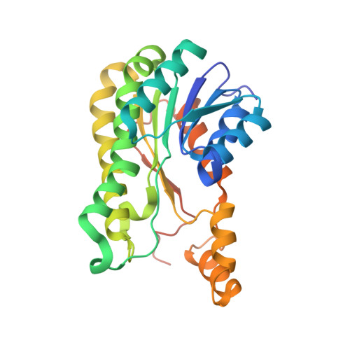
1YDE - PubMed Abstract:
To this day, a significant proportion of the human genome remains devoid of functional characterization. In this study, we present evidence that the previously functionally uncharacterized product of the human DHRS10 gene is endowed with 17beta-HSD (17beta-hydroxysteroid dehydrogenase) activity. 17beta-HSD enzymes are primarily involved in the metabolism of steroids at the C-17 position and also of other substrates such as fatty acids, prostaglandins and xenobiotics. In vitro, DHRS10 converts NAD+ into NADH in the presence of oestradiol, testosterone and 5-androstene-3beta,17beta-diol. Furthermore, the product of oestradiol oxidation, oestrone, was identified in intact cells transfected with a construct plasmid encoding the DHRS10 protein. In situ fluorescence hybridization studies have revealed the cytoplasmic localization of DHRS10. Along with tissue expression data, this suggests a role for DHRS10 in the local inactivation of steroids in the central nervous system and placenta. The crystal structure of the DHRS10 apoenzyme exhibits secondary structure of the SDR (short-chain dehydrogenase/reductase) family: a Rossmann-fold with variable loops surrounding the active site. It also reveals a broad and deep active site cleft into which NAD+ and oestradiol can be docked in a catalytically competent orientation.
Organizational Affiliation:
Structural Genomics Consortium, University of Oxford, Oxford OX3 7LD, UK. lukacikp@niddk.nih.gov