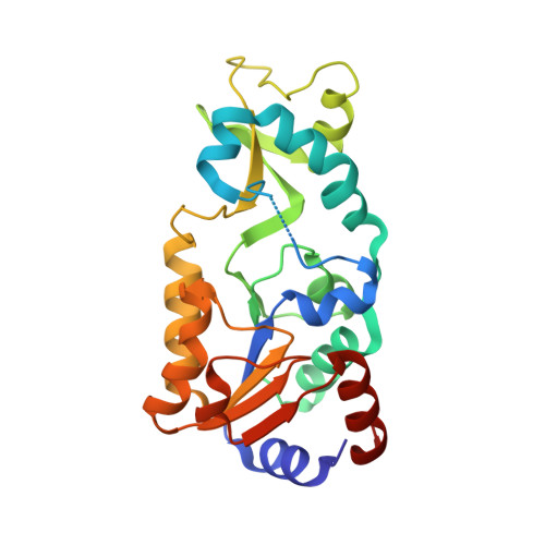
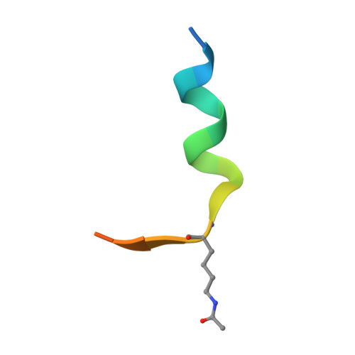
1YC5

Sir2-p53 peptide-nicotinamide


Experimental Data Snapshot

  • Method: X-RAY DIFFRACTION
  • Resolution: 1.40 Å
  • R-Value Free: 0.202 
  • R-Value Work: 0.185 
  • R-Value Observed: 0.186 

wwPDB Validation   3D Report Full Report


This is version 1.2 of the entry. See complete history


Literature

Mechanism of sirtuin inhibition by nicotinamide: altering the NAD(+) cosubstrate specificity of a Sir2 enzyme.

Avalos, J.L.Bever, K.M.Wolberger, C.

(2005) Mol Cell 17: 855-868

  • DOI: https://doi.org/10.1016/j.molcel.2005.02.022
  • Primary Citation of Related Structures:  
    1YC2, 1YC5

  • PubMed Abstract: 

    Sir2 enzymes form a unique class of NAD(+)-dependent deacetylases required for diverse biological processes, including transcriptional silencing, regulation of apoptosis, fat mobilization, and lifespan regulation. Sir2 activity is regulated by nicotinamide, a noncompetitive inhibitor that promotes a base-exchange reaction at the expense of deacetylation. To elucidate the mechanism of nicotinamide inhibition, we determined ternary complex structures of Sir2 enzymes containing nicotinamide. The structures show that free nicotinamide binds in a conserved pocket that participates in NAD(+) binding and catalysis. Based on our structures, we engineered a mutant that deacetylates peptides by using nicotinic acid adenine dinucleotide (NAAD) as a cosubstrate and is inhibited by nicotinic acid. The characteristics of the altered specificity enzyme establish that Sir2 enzymes contain a single site that participates in catalysis and nicotinamide regulation and provides additional insights into the Sir2 catalytic mechanism.


  • Organizational Affiliation

    Howard Hughes Medical Institute, Department of Biophysics and Biophysical Chemistry, School of Medicine, Johns Hopkins University, 725 N. Wolfe Street, Baltimore, Maryland 21205, USA.


Macromolecules
Find similar proteins by:  (by identity cutoff)  |  3D Structure
Entity ID: 1
MoleculeChains Sequence LengthOrganismDetailsImage
NAD-dependent deacetylase246Thermotoga maritimaMutation(s): 0 
Gene Names: npdA
EC: 3.5.1
UniProt
Find proteins for Q9WYW0 (Thermotoga maritima (strain ATCC 43589 / DSM 3109 / JCM 10099 / NBRC 100826 / MSB8))
Explore Q9WYW0 
Go to UniProtKB:  Q9WYW0
Entity Groups  
Sequence Clusters30% Identity50% Identity70% Identity90% Identity95% Identity100% Identity
UniProt GroupQ9WYW0
Sequence Annotations
Expand
  • Reference Sequence

Find similar proteins by:  Sequence   |   3D Structure  

Entity ID: 2
MoleculeChains Sequence LengthOrganismDetailsImage
Cellular tumor antigen p53 peptide18N/AMutation(s): 1 
UniProt & NIH Common Fund Data Resources
Find proteins for P04637 (Homo sapiens)
Explore P04637 
Go to UniProtKB:  P04637
PHAROS:  P04637
GTEx:  ENSG00000141510 
Entity Groups  
Sequence Clusters30% Identity50% Identity70% Identity90% Identity95% Identity100% Identity
UniProt GroupP04637
Sequence Annotations
Expand
  • Reference Sequence
Small Molecules
Modified Residues  1 Unique
IDChains TypeFormula2D DiagramParent
ALY
Query on ALY
B
L-PEPTIDE LINKINGC8 H16 N2 O3LYS
Binding Affinity Annotations 
IDSourceBinding Affinity
NCA PDBBind:  1YC5 IC50: 1.00e+6 (nM) from 1 assay(s)
Binding MOAD:  1YC5 IC50: 1.00e+6 (nM) from 1 assay(s)
Experimental Data & Validation

Experimental Data

  • Method: X-RAY DIFFRACTION
  • Resolution: 1.40 Å
  • R-Value Free: 0.202 
  • R-Value Work: 0.185 
  • R-Value Observed: 0.186 
  • Space Group: P 21 21 21
Unit Cell:
Length ( Å )Angle ( ˚ )
a = 46.1α = 90
b = 59.749β = 90
c = 106.121γ = 90
Software Package:
Software NamePurpose
REFMACrefinement
HKL-2000data reduction
SCALEPACKdata scaling
MOLREPphasing

Structure Validation

View Full Validation Report


Entry History 

Deposition Data

Revision History  (Full details and data files)

  • Version 1.0: 2005-04-26
    Type: Initial release
  • Version 1.1: 2008-04-30
    Changes: Version format compliance
  • Version 1.2: 2011-07-13
    Changes: Version format compliance