Crystal Structure of the Flagellar Rotor Protein FliN from Thermotoga maritima
Brown, P.N., Mathews, M.A.A., Joss, L.A., Hill, C.P., Blair, D.F.(2005) J Bacteriol 187: 2890-2902
- PubMed: 15805535
- DOI: https://doi.org/10.1128/JB.187.8.2890-2902.2005
- Primary Citation of Related Structures:
1YAB - PubMed Abstract:
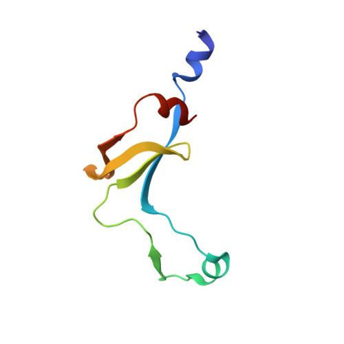
FliN is a component of the bacterial flagellum that is present at levels of more than 100 copies and forms the bulk of the C ring, a drum-shaped structure at the inner end of the basal body. FliN interacts with FliG and FliM to form the rotor-mounted switch complex that controls clockwise-counterclockwise switching of the motor. In addition to its functions in motor rotation and switching, FliN is thought to have a role in the export of proteins that form the exterior structures of the flagellum (the rod, hook, and filament). Here, we describe the crystal structure of most of the FliN protein of Thermotoga maritima. FliN is a tightly intertwined dimer composed mostly of beta sheet. Several well-conserved hydrophobic residues form a nonpolar patch on the surface of the molecule. A mutation in the hydrophobic patch affected both flagellar assembly and switching, showing that this surface feature is important for FliN function. The association state of FliN in solution was studied by analytical ultracentrifugation, which provided clues to the higher-level organization of the protein. T. maritima FliN is primarily a dimer in solution, and T. maritima FliN and FliM together form a stable FliM(1)-FliN(4) complex. Escherichia coli FliN forms a stable tetramer in solution. The arrangement of FliN subunits in the tetramer was modeled by reference to the crystal structure of tetrameric HrcQB(C), a related protein that functions in virulence factor secretion in Pseudomonas syringae. The modeled tetramer is elongated, with approximate dimensions of 110 by 40 by 35 Angstroms, and it has a large hydrophobic cleft formed from the hydrophobic patches on the dimers. On the basis of the present data and available electron microscopic images, we propose a model for the organization of FliN subunits in the C ring.
Organizational Affiliation:
Department of Biology, University of Utah, Salt Lake City, UT 84132, USA.