1Y3G

Crystal Structure of a Silanediol Protease Inhibitor Bound to Thermolysin


Experimental Data Snapshot

  • Method: X-RAY DIFFRACTION
  • Resolution: 2.10 Å
  • R-Value Free: 0.225 
  • R-Value Work: 0.155 
  • R-Value Observed: 0.158 

wwPDB Validation   3D Report Full Report


Ligand Structure Quality Assessment 


This is version 1.4 of the entry. See complete history


Literature

Structural Analysis of Silanediols as Transition-State-Analogue Inhibitors of the Benchmark Metalloprotease Thermolysin(,).

Juers, D.H.Kim, J.Matthews, B.W.Sieburth, S.M.

(2005) Biochemistry 44: 16524-16528

  • DOI: https://doi.org/10.1021/bi051346v
  • Primary Citation of Related Structures:  
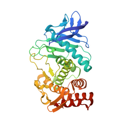
    1Y3G

  • PubMed Abstract: 

    Dialkylsilanediols have been found to be an effective functional group for the design of active-site-directed protease inhibitors, including aspartic (HIV protease) and metallo (ACE and thermolysin) proteases. The use of silanediols is predicated on its resemblance to the hydrated carbonyl transition-state structure of amide hydrolysis. This concept has been tested by replacing the presumed tetrahedral carbon of a thermolysin substrate with a silanediol group, resulting in an inhibitor with an inhibition constant K(i) = 40 nM. The structure of the silanediol bound to the active site of thermolysin was found to have a conformation very similar to that of a corresponding phosphonamidate inhibitor (K(i) = 10 nM). In both cases, a single oxygen is within bonding distance to the active-site zinc ion, mimicking the presumed tetrahedral transition state. There are binding differences that appear to be related to the presence or absence of protons on the oxygens attached to the silicon or phosphorus. This is the first crystal structure of an organosilane bound to the active site of a protease.


  • Organizational Affiliation

    Institute of Molecular Biology, Howard Hughes Medical Institute and Department of Physics, University of Oregon, Eugene, Oregon 97403, USA. juersdh@whitman.edu


Macromolecules
Find similar proteins by:  (by identity cutoff)  |  3D Structure
Entity ID: 1
MoleculeChains Sequence LengthOrganismDetailsImage
ThermolysinA [auth E]316Bacillus thermoproteolyticusMutation(s): 0 
EC: 3.4.24.27
UniProt
Find proteins for P00800 (Bacillus thermoproteolyticus)
Explore P00800 
Go to UniProtKB:  P00800
Entity Groups  
Sequence Clusters30% Identity50% Identity70% Identity90% Identity95% Identity100% Identity
UniProt GroupP00800
Sequence Annotations
Expand
  • Reference Sequence
Small Molecules
Ligands 6 Unique
IDChains Name / Formula / InChI Key2D Diagram3D Interactions
AMM
Query on AMM

Download Ideal Coordinates CCD File 
J [auth E](2S)-2-{[(AMINOMETHYL)(DIHYDROXY)SILYL]METHYL}-4-METHYLPENTANAL
C8 H19 N O3 Si
ZZKRIEGAOOCWBS-QMMMGPOBSA-N
3PL
Query on 3PL

Download Ideal Coordinates CCD File 
I [auth E]3-PHENYLPROPANAL
C9 H10 O
YGCZTXZTJXYWCO-UHFFFAOYSA-N
LEU
Query on LEU

Download Ideal Coordinates CCD File 
K [auth E]LEUCINE
C6 H13 N O2
ROHFNLRQFUQHCH-YFKPBYRVSA-N
DMS
Query on DMS

Download Ideal Coordinates CCD File 
G [auth E],
H [auth E]
DIMETHYL SULFOXIDE
C2 H6 O S
IAZDPXIOMUYVGZ-UHFFFAOYSA-N
ZN
Query on ZN

Download Ideal Coordinates CCD File 
F [auth E]ZINC ION
Zn
PTFCDOFLOPIGGS-UHFFFAOYSA-N
CA
Query on CA

Download Ideal Coordinates CCD File 
B [auth E],
C [auth E],
D [auth E],
E
CALCIUM ION
Ca
BHPQYMZQTOCNFJ-UHFFFAOYSA-N
Experimental Data & Validation

Experimental Data

  • Method: X-RAY DIFFRACTION
  • Resolution: 2.10 Å
  • R-Value Free: 0.225 
  • R-Value Work: 0.155 
  • R-Value Observed: 0.158 
  • Space Group: P 61 2 2
Unit Cell:
Length ( Å )Angle ( ˚ )
a = 93.52α = 90
b = 93.52β = 90
c = 131.77γ = 120
Software Package:
Software NamePurpose
TNTrefinement
PDB_EXTRACTdata extraction
CCP4data scaling
TNTphasing

Structure Validation

View Full Validation Report


Ligand Structure Quality Assessment 


Entry History 

Deposition Data

Revision History  (Full details and data files)

  • Version 1.0: 2006-01-17
    Type: Initial release
  • Version 1.1: 2008-04-30
    Changes: Version format compliance
  • Version 1.2: 2011-07-13
    Changes: Version format compliance
  • Version 1.3: 2017-10-11
    Changes: Refinement description
  • Version 1.4: 2024-02-14
    Changes: Data collection, Database references, Derived calculations