1Y2A

Structure of mammalian importin bound to the non-classical PLSCR1-NLS


Experimental Data Snapshot

  • Method: X-RAY DIFFRACTION
  • Resolution: 2.20 Å
  • R-Value Free: 0.258 
  • R-Value Work: 0.229 
  • R-Value Observed: 0.231 

wwPDB Validation   3D Report Full Report


This is version 1.3 of the entry. See complete history


Literature

Phospholipid Scramblase 1 Contains a Nonclassical Nuclear Localization Signal with Unique Binding Site in Importin alpha

Chen, M.-H.Ben-Efraim, I.Mitrousis, G.Walker-Kopp, N.Sims, P.J.Cingolani, G.

(2005) J Biol Chem 280: 10599-10606

  • DOI: https://doi.org/10.1074/jbc.M413194200
  • Primary Citation of Related Structures:  
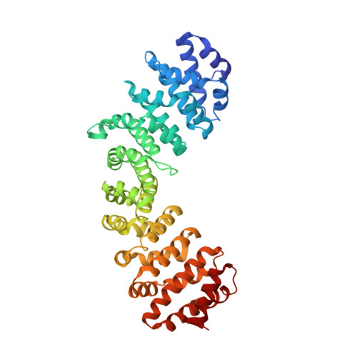
    1Y2A

  • PubMed Abstract: 

    Nuclear import of proteins containing a classical nuclear localization signal (NLS) is an energy-dependent process that requires the heterodimer importin alpha/beta. Three to six basic contiguous arginine/lysine residues characterize a classical NLS and are thought to form a basic patch on the surface of the import cargo. In this study, we have characterized the NLS of phospholipid scramblase 1 (PLSCR1), a lipid-binding protein that enters the nucleus via the nonclassical NLS (257)GKISKHWTGI(266). This import sequence lacks a contiguous stretch of positively charged residues, and it is enriched in hydrophobic residues. We have determined the 2.2 A crystal structure of a complex between the PLSCR1 NLS and the armadillo repeat core of vertebrate importin alpha. Our crystallographic analysis reveals that PLSCR1 NLS binds to armadillo repeats 1-4 of importin alpha, but its interaction partially overlaps the classical NLS binding site. Two PLSCR1 lysines occupy the canonical positions indicated as P2 and P5. Moreover, we present in vivo evidence that the critical lysine at position P2, which is essential in other known NLS sequences, is dispensable in PLSCR1 NLS. Taken together, these data provide insight into a novel nuclear localization signal that presents a distinct motif for binding to importin alpha.


  • Organizational Affiliation

    Department of Biochemistry and Molecular Biology, State University of New York Upstate Medical University, 750, E. Adams St., Syracuse, NY 13210, USA.


Macromolecules
Find similar proteins by:  (by identity cutoff)  |  3D Structure
Entity ID: 1
MoleculeChains Sequence LengthOrganismDetailsImage
Importin alpha-2 SubunitA [auth C]428Mus musculusMutation(s): 0 
Gene Names: Kpna2Rch1
UniProt & NIH Common Fund Data Resources
Find proteins for P52293 (Mus musculus)
Explore P52293 
Go to UniProtKB:  P52293
IMPC:  MGI:103561
Entity Groups  
Sequence Clusters30% Identity50% Identity70% Identity90% Identity95% Identity100% Identity
UniProt GroupP52293
Sequence Annotations
Expand
  • Reference Sequence

Find similar proteins by:  Sequence   |   3D Structure  

Entity ID: 2
MoleculeChains Sequence LengthOrganismDetailsImage
decamer fragment of Phospholipid scramblase 1B [auth P]10N/AMutation(s): 0 
UniProt & NIH Common Fund Data Resources
Find proteins for O15162 (Homo sapiens)
Explore O15162 
Go to UniProtKB:  O15162
PHAROS:  O15162
GTEx:  ENSG00000188313 
Entity Groups  
Sequence Clusters30% Identity50% Identity70% Identity90% Identity95% Identity100% Identity
UniProt GroupO15162
Sequence Annotations
Expand
  • Reference Sequence
Experimental Data & Validation

Experimental Data

  • Method: X-RAY DIFFRACTION
  • Resolution: 2.20 Å
  • R-Value Free: 0.258 
  • R-Value Work: 0.229 
  • R-Value Observed: 0.231 
  • Space Group: P 21 21 21
Unit Cell:
Length ( Å )Angle ( ˚ )
a = 78.239α = 90
b = 91.1β = 90
c = 97.247γ = 90
Software Package:
Software NamePurpose
DENZOdata reduction
SCALEPACKdata scaling
CNSrefinement
CNSphasing

Structure Validation

View Full Validation Report


Entry History 

Deposition Data

Revision History  (Full details and data files)

  • Version 1.0: 2005-02-01
    Type: Initial release
  • Version 1.1: 2008-04-30
    Changes: Version format compliance
  • Version 1.2: 2011-07-13
    Changes: Version format compliance
  • Version 1.3: 2024-02-14
    Changes: Data collection, Database references