


Crystal structure of of the SH3 domain of phospholipase C Gamma-1
Mariuzza, R., Sangwoo, C.To be published.
Experimental Data Snapshot
wwPDB Validation 3D Report Full Report

Entity ID: 1 | |||||
|---|---|---|---|---|---|
| Molecule | Chains | Sequence Length | Organism | Details | Image |
| 1-phosphatidylinositol-4,5-bisphosphate phosphodiesterase gamma 1 | 61 | Rattus norvegicus | Mutation(s): 1 Gene Names: Plcg1 EC: 3.1.4.11 | 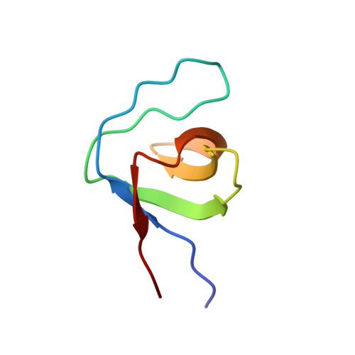 | |
UniProt | |||||
Find proteins for P10686 (Rattus norvegicus) Explore P10686 Go to UniProtKB: P10686 | |||||
Entity Groups | |||||
| Sequence Clusters | 30% Identity50% Identity70% Identity90% Identity95% Identity100% Identity | ||||
| UniProt Group | P10686 | ||||
Sequence AnnotationsExpand | |||||
| |||||
| Length ( Å ) | Angle ( ˚ ) |
|---|---|
| a = 28.748 | α = 90 |
| b = 30.978 | β = 92.12 |
| c = 29.753 | γ = 90 |
| Software Name | Purpose |
|---|---|
| DENZO | data reduction |
| SCALEPACK | data scaling |
| MOLREP | phasing |
| REFMAC | refinement |
| PDB_EXTRACT | data extraction |
RCSB PDB (citation) is hosted by
RCSB PDB is a member of the

