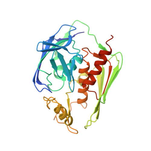
Refined Solution Structure of the LpxC-TU-514 Complex and pK(a) Analysis of an Active Site Histidine: Insights into the Mechanism and Inhibitor Design
Coggins, B.E., McClerren, A.L., Jiang, L., Li, X., Rudolph, J., Hindsgaul, O., Raetz, C.R.H., Zhou, P.(2005) Biochemistry 44: 1114-1126
- PubMed: 15667205
- DOI: https://doi.org/10.1021/bi047820z
- Primary Citation of Related Structures:
1XXE - PubMed Abstract:
Lipopolysaccharide, the major constituent of the outer monolayer of the outer membrane of Gram-negative bacteria, is anchored into the membrane through the hydrophobic moiety lipid A, a hexaacylated disaccharide. The zinc-dependent metalloamidase UDP-3-O-acyl-N-acetylglucosamine deacetylase (LpxC) catalyzes the second and committed step in the biosynthesis of lipid A. LpxC shows no homology to mammalian metalloamidases and is essential for cell viability, making it an important target for the development of novel antibacterial compounds. Recent NMR and X-ray studies of the LpxC from Aquifex aeolicus have provided the first structural information about this family of proteins. Insight into the catalytic mechanism and the design of effective inhibitors could be facilitated by more detailed structural and biochemical studies that define substrate-protein interactions and the roles of specific residues in the active site. Here, we report the synthesis of the (13)C-labeled substrate-analogue inhibitor TU-514, and the subsequent refinement of the solution structure of the A. aeolicus LpxC-TU-514 complex using residual dipolar couplings. We also reevaluate the catalytic role of an active site histidine, H253, on the basis of both its pK(a) as determined by NMR titration and pH-dependent kinetic analyses. These results provide a structural basis for the design of more potent LpxC inhibitors than those that are currently available.
Organizational Affiliation:
Department of Biochemistry, Duke University Medical Center, P.O. Box 3711, 242 Nanaline Duke Building, Research Drive, Durham, North Carolina 27710, USA.


