1XVX

Crystal Structure of iron-loaded Yersinia enterocolitica YfuA


Experimental Data Snapshot

  • Method: X-RAY DIFFRACTION
  • Resolution: 1.53 Å
  • R-Value Free: 0.193 
  • R-Value Work: 0.161 
  • R-Value Observed: 0.163 

wwPDB Validation   3D Report Full Report


This is version 1.3 of the entry. See complete history


Literature

Novel Anion-independent Iron Coordination by Members of a Third Class of Bacterial Periplasmic Ferric Ion-binding Proteins

Shouldice, S.R.McRee, D.E.Dougan, D.R.Tari, L.W.Schryvers, A.B.

(2005) J Biol Chem 280: 5820-5827

  • DOI: https://doi.org/10.1074/jbc.M411238200
  • Primary Citation of Related Structures:  
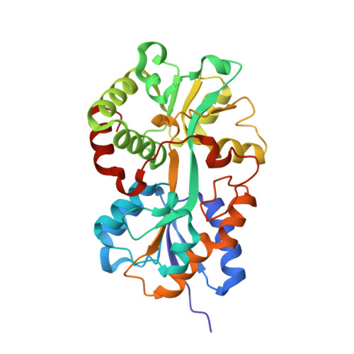
    1XVX, 1XVY

  • PubMed Abstract: 

    The uptake of the element iron is vital for the survival of most organisms. Numerous pathogenic Gram-negative bacteria utilize a periplasm-to-cytosol ATP-binding cassette transport pathway to transport this essential atom in to the cell. In this study, we investigated the Yersinia enterocolitica (YfuA) and Serratia marcescens (SfuA) iron-binding periplasmic proteins. We have determined the 1.8-angstroms structures of iron-loaded (YfuA) and iron-free (SfuA) forms of this class of proteins. Although the sequence of these proteins varies considerably from the other members of the transferrin structural superfamily, they adopt the same three-dimensional fold. The iron-loaded YfuA structure illustrates the unique nature of this new class of proteins in that they are able to octahedrally coordinate the ferric ion in the absence of a bound anion. The iron-free SfuA structure contains a bound citrate anion in the iron-binding cleft that tethers the N- and C-terminal domains of the apo protein and stabilizes the partially open structure.


  • Organizational Affiliation

    Department of Microbiology and Infectious Diseases, University of Calgary, Calgary, Alberta T2N 4N1, Canada.


Macromolecules
Find similar proteins by:  (by identity cutoff)  |  3D Structure
Entity ID: 1
MoleculeChains Sequence LengthOrganismDetailsImage
YfuA312Yersinia enterocoliticaMutation(s): 0 
Gene Names: yfuA
UniProt
Find proteins for A1JLH5 (Yersinia enterocolitica serotype O:8 / biotype 1B (strain NCTC 13174 / 8081))
Explore A1JLH5 
Go to UniProtKB:  A1JLH5
Entity Groups  
Sequence Clusters30% Identity50% Identity70% Identity90% Identity95% Identity100% Identity
UniProt GroupA1JLH5
Sequence Annotations
Expand
  • Reference Sequence
Small Molecules
Ligands 4 Unique
IDChains Name / Formula / InChI Key2D Diagram3D Interactions
ZN
Query on ZN

Download Ideal Coordinates CCD File 
C [auth A],
D [auth A],
E [auth A],
F [auth A],
G [auth A]
ZINC ION
Zn
PTFCDOFLOPIGGS-UHFFFAOYSA-N
EDO
Query on EDO

Download Ideal Coordinates CCD File 
I [auth A],
J [auth A]
1,2-ETHANEDIOL
C2 H6 O2
LYCAIKOWRPUZTN-UHFFFAOYSA-N
CO3
Query on CO3

Download Ideal Coordinates CCD File 
H [auth A]CARBONATE ION
C O3
BVKZGUZCCUSVTD-UHFFFAOYSA-L
FE
Query on FE

Download Ideal Coordinates CCD File 
B [auth A]FE (III) ION
Fe
VTLYFUHAOXGGBS-UHFFFAOYSA-N
Experimental Data & Validation

Experimental Data

  • Method: X-RAY DIFFRACTION
  • Resolution: 1.53 Å
  • R-Value Free: 0.193 
  • R-Value Work: 0.161 
  • R-Value Observed: 0.163 
  • Space Group: C 1 2 1
Unit Cell:
Length ( Å )Angle ( ˚ )
a = 108.392α = 90
b = 46.951β = 103.51
c = 62.454γ = 90
Software Package:
Software NamePurpose
REFMACrefinement
d*TREKdata reduction
CCP4data scaling
SHELXDphasing
SHELXEmodel building

Structure Validation

View Full Validation Report


Entry History 

Deposition Data

Revision History  (Full details and data files)

  • Version 1.0: 2004-12-14
    Type: Initial release
  • Version 1.1: 2008-04-30
    Changes: Version format compliance
  • Version 1.2: 2011-07-13
    Changes: Advisory, Derived calculations, Version format compliance
  • Version 1.3: 2024-02-14
    Changes: Data collection, Database references, Derived calculations