1XKY

Crystal Structure of Dihydrodipicolinate Synthase DapA-2 (BA3935) from Bacillus Anthracis at 1.94A Resolution.


Experimental Data Snapshot

  • Method: X-RAY DIFFRACTION
  • Resolution: 1.94 Å
  • R-Value Free: 0.197 
  • R-Value Work: 0.153 
  • R-Value Observed: 0.156 

wwPDB Validation   3D Report Full Report


This is version 1.3 of the entry. See complete history


Literature

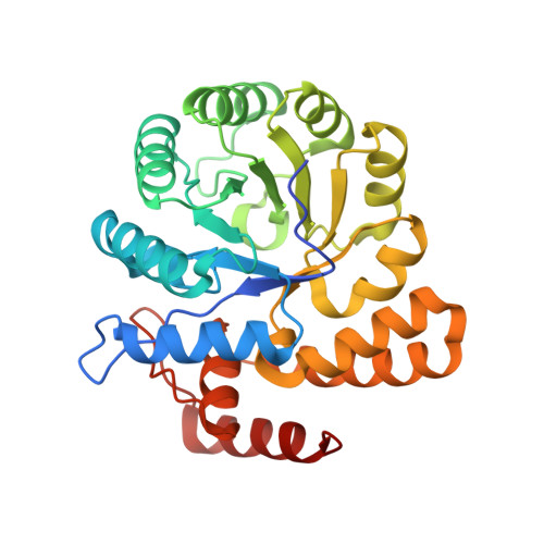
Crystal structure of dihydrodipicolinate synthase (BA3935) from Bacillus anthracis at 1.94 A resolution

Blagova, E.Levdikov, V.Milioti, N.Fogg, M.J.Kalliomaa, A.K.Brannigan, J.A.Wilson, K.S.Wilkinson, A.J.

(2006) Proteins 62: 297-301


Macromolecules
Find similar proteins by:  (by identity cutoff)  |  3D Structure
Entity ID: 1
MoleculeChains Sequence LengthOrganismDetailsImage
dihydrodipicolinate synthase
A, B, C, D
301Bacillus anthracisMutation(s): 0 
Gene Names: DapA-2
EC: 4.2.1.52
UniProt
Find proteins for A0A6L7H340 (Bacillus anthracis)
Explore A0A6L7H340 
Go to UniProtKB:  A0A6L7H340
Entity Groups  
Sequence Clusters30% Identity50% Identity70% Identity90% Identity95% Identity100% Identity
UniProt GroupA0A6L7H340
Sequence Annotations
Expand
  • Reference Sequence
Experimental Data & Validation

Experimental Data

  • Method: X-RAY DIFFRACTION
  • Resolution: 1.94 Å
  • R-Value Free: 0.197 
  • R-Value Work: 0.153 
  • R-Value Observed: 0.156 
  • Space Group: P 21 21 21
Unit Cell:
Length ( Å )Angle ( ˚ )
a = 55.203α = 90
b = 126.882β = 90
c = 173.319γ = 90
Software Package:
Software NamePurpose
REFMACrefinement
DENZOdata reduction
SCALEPACKdata scaling
MOLREPphasing

Structure Validation

View Full Validation Report


Entry History 

Deposition Data

Revision History  (Full details and data files)

  • Version 1.0: 2005-10-04
    Type: Initial release
  • Version 1.1: 2008-04-30
    Changes: Version format compliance
  • Version 1.2: 2011-07-13
    Changes: Version format compliance
  • Version 1.3: 2023-08-23
    Changes: Data collection, Database references, Derived calculations, Refinement description