


Crystal structure of Nucleoside diphosphate kinase B from Plasmodium falciparum
Robien, M.A., Bosch, J., Hol, W.G.J., Structural Genomics of Pathogenic Protozoa ConsortiumTo be published.
Experimental Data Snapshot
wwPDB Validation 3D Report Full Report

Entity ID: 1 | |||||
|---|---|---|---|---|---|
| Molecule | Chains | Sequence Length | Organism | Details | Image |
| Nucleoside diphosphate kinase B | 157 | Plasmodium falciparum 3D7 | Mutation(s): 0 Gene Names: PF13_0349 EC: 2.7.4.6 | 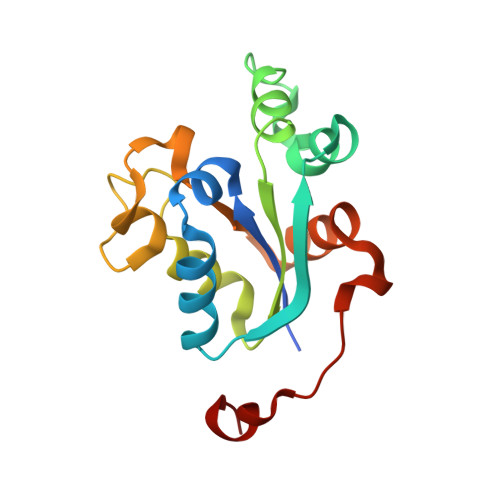 | |
UniProt | |||||
Find proteins for Q8ID43 (Plasmodium falciparum (isolate 3D7)) Explore Q8ID43 Go to UniProtKB: Q8ID43 | |||||
Entity Groups | |||||
| Sequence Clusters | 30% Identity50% Identity70% Identity90% Identity95% Identity100% Identity | ||||
| UniProt Group | Q8ID43 | ||||
Sequence AnnotationsExpand | |||||
| |||||
| Length ( Å ) | Angle ( ˚ ) |
|---|---|
| a = 68.593 | α = 90 |
| b = 112.424 | β = 119.05 |
| c = 68.59 | γ = 90 |
| Software Name | Purpose |
|---|---|
| REFMAC | refinement |
| MOSFLM | data reduction |
| CCP4 | data scaling |
| SHARP | phasing |
RCSB PDB (citation) is hosted by
RCSB PDB is a member of the

