1XHU

HincII bound to cleaved, cognate DNA containing GTCGAC


Experimental Data Snapshot

  • Method: X-RAY DIFFRACTION
  • Resolution: 2.95 Å
  • R-Value Free: 0.277 
  • R-Value Work: 0.187 

wwPDB Validation   3D Report Full Report


This is version 1.4 of the entry. See complete history


Literature

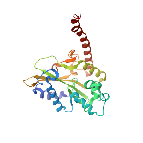
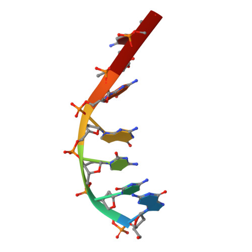
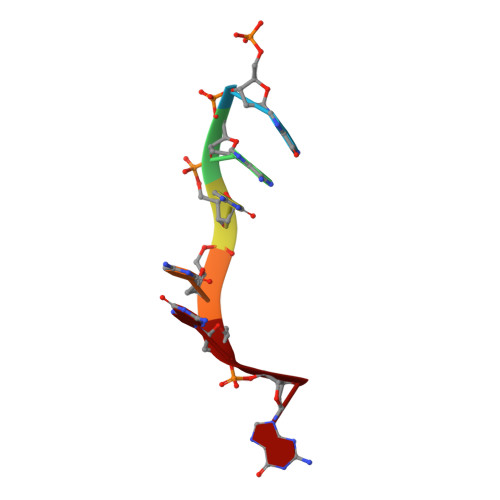
Mechanistic Insights from the Structures of HincII Bound to Cognate DNA Cleaved from Addition of Mg(2+) and Mn(2+)

Etzkorn, C.Horton, N.C.

(2004) J Mol Biol 343: 833-849

  • DOI: https://doi.org/10.1016/j.jmb.2004.08.082
  • Primary Citation of Related Structures:  
    1XHU, 1XHV

  • PubMed Abstract: 

    The three-dimensional X-ray crystal structures of HincII bound to cognate DNA containing GTCGAC and Mn(2+) or Mg(2+), at 2.50A and 2.95A resolution, respectively, are presented. In both structures, the DNA is found cleaved, and the positions of the active-site groups, cleaved phosphate group, and 3' oxygen atom of the leaving group are in very similar positions. Two highly occupied Mn(2+) positions are found in each active site of the four crystallographically independent subunit copies in the HincII/DNA/Mn(2+) structure. The manganese ion closest to the previously identified single Ca(2+) position of HincII is shifted 1.7A and has lost direct ligation to the active-site aspartate residue, Asp127. A Mn(2+)-ligated water molecule in a position analogous to that seen in the HincII/DNA/Ca(2+) structure, and proposed to be the attacking nucleophile, is beyond hydrogen bonding distance from the active-site lysine residue, Lys129, but remains within hydrogen bonding distance from the proRp oxygen atom of the phosphate group 3' to the scissile phosphate group. In addition, the position of the cleaved phosphate group is on the opposite side of the axis connecting the two metal ions relative to that found in the BamHI/product DNA/Mn(2+) structure. Mechanistic implications are discussed, and a model for the two-metal-ion mechanism of DNA cleavage by HincII is proposed.


  • Organizational Affiliation

    Department of Biochemistry and Molecular Biophysics, University of Arizona, Tucson, AZ 85721, USA.


Macromolecules

Find similar proteins by:  (by identity cutoff)  |  3D Structure
Entity ID: 3
MoleculeChains Sequence LengthOrganismDetailsImage
Type II restriction enzyme HincIII [auth A],
J [auth B],
K [auth C],
L [auth D]
257Haemophilus influenzaeMutation(s): 0 
Gene Names: hincIIR
EC: 3.1.21.4
UniProt
Find proteins for P17743 (Haemophilus influenzae)
Explore P17743 
Go to UniProtKB:  P17743
Entity Groups  
Sequence Clusters30% Identity50% Identity70% Identity90% Identity95% Identity100% Identity
UniProt GroupP17743
Sequence Annotations
Expand
  • Reference Sequence

Find similar nucleic acids by:  Sequence   |   3D Structure  

Entity ID: 1
MoleculeChains LengthOrganismImage
5'-D(*GP*CP*CP*GP*GP*TP*C)-3'A [auth E],
C [auth G],
E [auth I],
G [auth K]
7N/A
Sequence Annotations
Expand
  • Reference Sequence

Find similar nucleic acids by:  Sequence   |   3D Structure  

Entity ID: 2
MoleculeChains LengthOrganismImage
5'-D(P*GP*AP*CP*CP*GP*G)-3'B [auth F],
D [auth H],
F [auth J],
H [auth L]
6N/A
Sequence Annotations
Expand
  • Reference Sequence
Experimental Data & Validation

Experimental Data

  • Method: X-RAY DIFFRACTION
  • Resolution: 2.95 Å
  • R-Value Free: 0.277 
  • R-Value Work: 0.187 
  • Space Group: I 21 21 21
Unit Cell:
Length ( Å )Angle ( ˚ )
a = 67.2α = 90
b = 177.2β = 90
c = 256γ = 90
Software Package:
Software NamePurpose
XDSdata scaling
SCALAdata scaling
CNSrefinement
XDSdata reduction
CCP4data scaling
CNSphasing

Structure Validation

View Full Validation Report


Entry History 

Deposition Data

Revision History  (Full details and data files)

  • Version 1.0: 2004-09-28
    Type: Initial release
  • Version 1.1: 2008-04-30
    Changes: Version format compliance
  • Version 1.2: 2011-07-13
    Changes: Version format compliance
  • Version 1.3: 2017-10-11
    Changes: Refinement description
  • Version 1.4: 2023-08-23
    Changes: Data collection, Database references, Refinement description