1XG4

Crystal Structure of the C123S 2-Methylisocitrate Lyase Mutant from Escherichia coli in complex with the inhibitor isocitrate


Experimental Data Snapshot

  • Method: X-RAY DIFFRACTION
  • Resolution: 1.60 Å
  • R-Value Free: 0.201 
  • R-Value Work: 0.181 

wwPDB Validation   3D Report Full Report


Ligand Structure Quality Assessment 


This is version 1.4 of the entry. See complete history


Literature

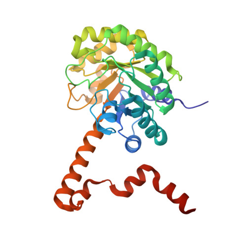
Crystal Structures of 2-Methylisocitrate Lyase in Complex with Product and with Isocitrate Inhibitor Provide Insight into Lyase Substrate Specificity, Catalysis and Evolution

Liu, S.Lu, Z.Han, Y.Melamud, E.Dunaway-Mariano, D.Herzberg, O.

(2005) Biochemistry 44: 2949-2962

  • DOI: https://doi.org/10.1021/bi0479712
  • Primary Citation of Related Structures:  
    1OQF, 1XG3, 1XG4

  • PubMed Abstract: 

    Two crystal structures of the C123S mutant of 2-methylisocitrate lyase have been determined, one with the bound reaction products, Mg(2+)-pyruvate and succinate, and the second with a bound Mg(2+)-(2R,3S)-isocitrate inhibitor. Comparison with the structure of the wild-type enzyme in the unbound state reveals that the enzyme undergoes a conformational transition that sequesters the ligand from solvent, as previously observed for two other enzyme superfamily members, isocitrate lyase and phosphoenolpyruvate mutase. The binding modes reveal the determinants of substrate specificity and stereoselectivity, and the stringent specificity is verified in solution using various potential substrates. A model of bound 2-methylisocitrate has been developed based on the experimentally determined structures. We propose a catalytic mechanism involving an alpha-carboxy-carbanion intermediate/transition state, which is consistent with previous stereochemical experiments showing inversion of configuration at the C(3) of 2-methylisocitrate. Structure-based sequence analysis and phylogenic tree construction reveal determinants of substrate specificity, highlight nodes of divergence of families, and predict enzyme families with new functions.


  • Organizational Affiliation

    Center for Advanced Research in Biotechnology, University of Maryland Biotechnology Institute, Rockville, Maryland 20850, USA.


Macromolecules
Find similar proteins by:  (by identity cutoff)  |  3D Structure
Entity ID: 1
MoleculeChains Sequence LengthOrganismDetailsImage
Probable methylisocitrate lyase
A, B, C, D
295Escherichia coliMutation(s): 1 
Gene Names: prpB
EC: 4.1.3.30
UniProt
Find proteins for P77541 (Escherichia coli (strain K12))
Explore P77541 
Go to UniProtKB:  P77541
Entity Groups  
Sequence Clusters30% Identity50% Identity70% Identity90% Identity95% Identity100% Identity
UniProt GroupP77541
Sequence Annotations
Expand
  • Reference Sequence
Experimental Data & Validation

Experimental Data

  • Method: X-RAY DIFFRACTION
  • Resolution: 1.60 Å
  • R-Value Free: 0.201 
  • R-Value Work: 0.181 
  • Space Group: C 1 2 1
Unit Cell:
Length ( Å )Angle ( ˚ )
a = 159.061α = 90
b = 84.561β = 108.15
c = 99.756γ = 90
Software Package:
Software NamePurpose
DENZOdata reduction
SCALEPACKdata scaling
AMoREphasing
CNSrefinement

Structure Validation

View Full Validation Report


Ligand Structure Quality Assessment 


Entry History 

Deposition Data

Revision History  (Full details and data files)

  • Version 1.0: 2005-03-01
    Type: Initial release
  • Version 1.1: 2008-04-30
    Changes: Version format compliance
  • Version 1.2: 2011-07-13
    Changes: Version format compliance
  • Version 1.3: 2021-10-20
    Changes: Database references, Derived calculations
  • Version 1.4: 2023-08-23
    Changes: Data collection, Refinement description