1XFD

Structure of a human A-type Potassium Channel Accelerating factor DPPX, a member of the dipeptidyl aminopeptidase family


Experimental Data Snapshot

  • Method: X-RAY DIFFRACTION
  • Resolution: 3.00 Å
  • R-Value Free: 0.280 
  • R-Value Work: 0.248 
  • R-Value Observed: 0.248 

wwPDB Validation   3D Report Full Report


This is version 2.1 of the entry. See complete history


Literature

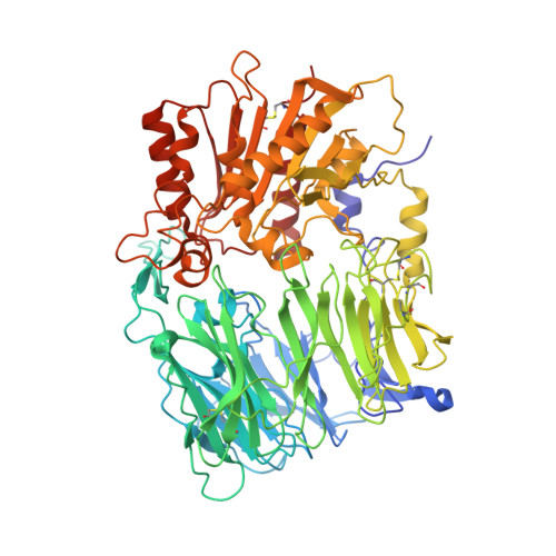
Structure of a human A-type potassium channel interacting protein DPPX, a member of the dipeptidyl aminopeptidase family

Strop, P.Bankovich, A.J.Hansen, K.C.Garcia, K.C.Brunger, A.T.

(2004) J Mol Biol 343: 1055-1065

  • DOI: https://doi.org/10.1016/j.jmb.2004.09.003
  • Primary Citation of Related Structures:  
    1XFD

  • PubMed Abstract: 

    It has recently been reported that dipeptidyl aminopeptidase X (DPPX) interacts with the voltage-gated potassium channel Kv4 and that co-expression of DPPX together with Kv4 pore forming alpha-subunits, and potassium channel interacting proteins (KChIPs), reconstitutes properties of native A-type potassium channels in vitro. Here we report the X-ray crystal structure of the extracellular domain of human DPPX determined at 3.0A resolution. This structure reveals the potential for a surface electrostatic change based on the protonation state of histidine. Subtle changes in extracellular pH might modulate the interaction of DPPX with Kv4.2 and possibly with other proteins. We propose models of DPPX interaction with the voltage-gated potassium channel complex. The dimeric structure of DPPX is highly homologous to the related protein DPP-IV. Comparison of the active sites of DPPX and DPP-IV reveals loss of the catalytic serine residue but the presence of an additional serine near the "active" site. However, the arrangement of residues is inconsistent with that of canonical serine proteases and DPPX is unlikely to function as a protease (dipeptidyl aminopeptidase).


  • Organizational Affiliation

    Howard Hughes Medical Institute and Department of Molecular and Cellular Physiology, Stanford Synchrotron Radiation Laboratory, Stanford University, James H. Clark Center E300, 318 Campus Drive, Stanford, CA 94305, USA.


Macromolecules
Find similar proteins by:  (by identity cutoff)  |  3D Structure
Entity ID: 1
MoleculeChains Sequence LengthOrganismDetailsImage
Dipeptidyl aminopeptidase-like protein 6
A, B, C, D
723Homo sapiensMutation(s): 0 
Gene Names: DPPX
UniProt & NIH Common Fund Data Resources
Find proteins for P42658 (Homo sapiens)
Explore P42658 
Go to UniProtKB:  P42658
PHAROS:  P42658
GTEx:  ENSG00000130226 
Entity Groups  
Sequence Clusters30% Identity50% Identity70% Identity90% Identity95% Identity100% Identity
UniProt GroupP42658
Sequence Annotations
Expand
  • Reference Sequence
Oligosaccharides

Help

Entity ID: 2
MoleculeChains Length2D Diagram Glycosylation3D Interactions
beta-D-mannopyranose-(1-4)-2-acetamido-2-deoxy-beta-D-glucopyranose-(1-4)-2-acetamido-2-deoxy-beta-D-glucopyranose
E, G, I, K, N
E, G, I, K, N, Q, T, V, Y, Z
3N-Glycosylation
Glycosylation Resources
GlyTouCan:  G15407YE
GlyCosmos:  G15407YE
GlyGen:  G15407YE
Entity ID: 3
MoleculeChains Length2D Diagram Glycosylation3D Interactions
beta-D-mannopyranose-(1-4)-alpha-D-mannopyranose-(1-4)-2-acetamido-2-deoxy-alpha-D-glucopyranose-(1-4)-2-acetamido-2-deoxy-beta-D-glucopyranose
F
4N-Glycosylation
Glycosylation Resources
GlyTouCan:  G63308WB
GlyCosmos:  G63308WB
GlyGen:  G63308WB
Entity ID: 4
MoleculeChains Length2D Diagram Glycosylation3D Interactions
alpha-D-mannopyranose-(1-4)-beta-D-mannopyranose-(1-4)-2-acetamido-2-deoxy-beta-D-glucopyranose-(1-4)-2-acetamido-2-deoxy-beta-D-glucopyranose
H
4N-Glycosylation
Glycosylation Resources
GlyTouCan:  G08918EL
GlyCosmos:  G08918EL
GlyGen:  G08918EL
Entity ID: 5
MoleculeChains Length2D Diagram Glycosylation3D Interactions
2-acetamido-2-deoxy-beta-D-glucopyranose-(1-4)-2-acetamido-2-deoxy-beta-D-glucopyranose
J, M, O, P, S
J, M, O, P, S, U
2N-Glycosylation
Glycosylation Resources
GlyTouCan:  G42666HT
GlyCosmos:  G42666HT
GlyGen:  G42666HT
Entity ID: 6
MoleculeChains Length2D Diagram Glycosylation3D Interactions
alpha-D-mannopyranose-(1-4)-2-acetamido-2-deoxy-alpha-D-glucopyranose-(1-4)-2-acetamido-2-deoxy-beta-D-glucopyranose
L, R, W
3N-Glycosylation
Glycosylation Resources
GlyTouCan:  G52260VY
GlyCosmos:  G52260VY
GlyGen:  G52260VY
Entity ID: 7
MoleculeChains Length2D Diagram Glycosylation3D Interactions
alpha-D-mannopyranose-(1-4)-alpha-D-mannopyranose-(1-4)-2-acetamido-2-deoxy-beta-D-glucopyranose-(1-4)-2-acetamido-2-deoxy-beta-D-glucopyranose
X
4N-Glycosylation
Glycosylation Resources
GlyTouCan:  G81874LD
GlyCosmos:  G81874LD
GlyGen:  G81874LD
Experimental Data & Validation

Experimental Data

  • Method: X-RAY DIFFRACTION
  • Resolution: 3.00 Å
  • R-Value Free: 0.280 
  • R-Value Work: 0.248 
  • R-Value Observed: 0.248 
  • Space Group: P 1 21 1
Unit Cell:
Length ( Å )Angle ( ˚ )
a = 68.98α = 90
b = 170.23β = 92.11
c = 159.316γ = 90
Software Package:
Software NamePurpose
DENZOdata reduction
SCALEPACKdata scaling
AMoREphasing
CNSrefinement

Structure Validation

View Full Validation Report


Entry History 

Deposition Data

Revision History  (Full details and data files)

  • Version 1.0: 2004-10-26
    Type: Initial release
  • Version 1.1: 2008-04-30
    Changes: Version format compliance
  • Version 1.2: 2011-07-13
    Changes: Non-polymer description, Version format compliance
  • Version 2.0: 2020-07-29
    Type: Remediation
    Reason: Carbohydrate remediation
    Changes: Advisory, Atomic model, Data collection, Derived calculations, Structure summary
  • Version 2.1: 2023-08-23
    Changes: Data collection, Database references, Refinement description, Structure summary