1XF9

Structure of NBD1 from murine CFTR- F508S mutant


Experimental Data Snapshot

  • Method: X-RAY DIFFRACTION
  • Resolution: 2.70 Å
  • R-Value Free: 0.253 
  • R-Value Work: 0.201 
  • R-Value Observed: 0.201 

wwPDB Validation   3D Report Full Report


Ligand Structure Quality Assessment 


This is version 1.4 of the entry. See complete history


Literature

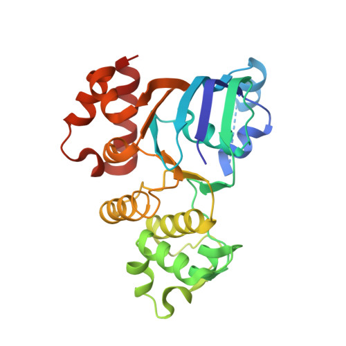
Side chain and backbone contributions of Phe508 to CFTR folding.

Thibodeau, P.H.Brautigam, C.A.Machius, M.Thomas, P.J.

(2005) Nat Struct Mol Biol 12: 10-16

  • DOI: https://doi.org/10.1038/nsmb881
  • Primary Citation of Related Structures:  
    1XF9, 1XFA

  • PubMed Abstract: 

    Mutations in the cystic fibrosis transmembrane conductance regulator (CFTR), an integral membrane protein, cause cystic fibrosis (CF). The most common CF-causing mutant, deletion of Phe508, fails to properly fold. To elucidate the role Phe508 plays in the folding of CFTR, missense mutations at this position were generated. Only one missense mutation had a pronounced effect on the stability and folding of the isolated domain in vitro. In contrast, many substitutions, including those of charged and bulky residues, disrupted folding of full-length CFTR in cells. Structures of two mutant nucleotide-binding domains (NBDs) reveal only local alterations of the surface near position 508. These results suggest that the peptide backbone plays a role in the proper folding of the domain, whereas the side chain plays a role in defining a surface of NBD1 that potentially interacts with other domains during the maturation of intact CFTR.


  • Organizational Affiliation

    Department of Physiology, The University of Texas Southwestern Medical Center at Dallas, 75390 USA.


Macromolecules
Find similar proteins by:  (by identity cutoff)  |  3D Structure
Entity ID: 1
MoleculeChains Sequence LengthOrganismDetailsImage
Cystic fibrosis transmembrane conductance regulator
A, B, C, D
283Mus musculusMutation(s): 1 
Gene Names: CftrAbcc7
UniProt
Find proteins for P26361 (Mus musculus)
Explore P26361 
Go to UniProtKB:  P26361
Entity Groups  
Sequence Clusters30% Identity50% Identity70% Identity90% Identity95% Identity100% Identity
UniProt GroupP26361
Sequence Annotations
Expand
  • Reference Sequence
Small Molecules
Ligands 3 Unique
IDChains Name / Formula / InChI Key2D Diagram3D Interactions
ATP
Query on ATP

Download Ideal Coordinates CCD File 
F [auth A],
K [auth B],
O [auth C],
S [auth D]
ADENOSINE-5'-TRIPHOSPHATE
C10 H16 N5 O13 P3
ZKHQWZAMYRWXGA-KQYNXXCUSA-N
ACY
Query on ACY

Download Ideal Coordinates CCD File 
G [auth A]
H [auth A]
L [auth B]
P [auth C]
T [auth D]
G [auth A],
H [auth A],
L [auth B],
P [auth C],
T [auth D],
U [auth D]
ACETIC ACID
C2 H4 O2
QTBSBXVTEAMEQO-UHFFFAOYSA-N
MG
Query on MG

Download Ideal Coordinates CCD File 
E [auth A]
I [auth B]
J [auth B]
M [auth C]
N [auth C]
E [auth A],
I [auth B],
J [auth B],
M [auth C],
N [auth C],
Q [auth D],
R [auth D]
MAGNESIUM ION
Mg
JLVVSXFLKOJNIY-UHFFFAOYSA-N
Experimental Data & Validation

Experimental Data

  • Method: X-RAY DIFFRACTION
  • Resolution: 2.70 Å
  • R-Value Free: 0.253 
  • R-Value Work: 0.201 
  • R-Value Observed: 0.201 
  • Space Group: P 4 21 2
Unit Cell:
Length ( Å )Angle ( ˚ )
a = 170.449α = 90
b = 170.449β = 90
c = 109.065γ = 90
Software Package:
Software NamePurpose
CNSrefinement
HKL-2000data reduction
SCALEPACKdata scaling
CNSphasing

Structure Validation

View Full Validation Report


Ligand Structure Quality Assessment 


Entry History 

Deposition Data

Revision History  (Full details and data files)

  • Version 1.0: 2004-12-28
    Type: Initial release
  • Version 1.1: 2008-04-30
    Changes: Version format compliance
  • Version 1.2: 2011-07-13
    Changes: Derived calculations, Version format compliance
  • Version 1.3: 2021-10-20
    Changes: Database references, Derived calculations
  • Version 1.4: 2023-08-23
    Changes: Data collection, Refinement description