1XEU

Crystal Structure of Internalin C from Listeria monocytogenes


Experimental Data Snapshot

  • Method: X-RAY DIFFRACTION
  • Resolution: 2.05 Å
  • R-Value Free: 0.243 
  • R-Value Work: 0.200 
  • R-Value Observed: 0.202 

wwPDB Validation   3D Report Full Report


This is version 1.4 of the entry. See complete history


Literature

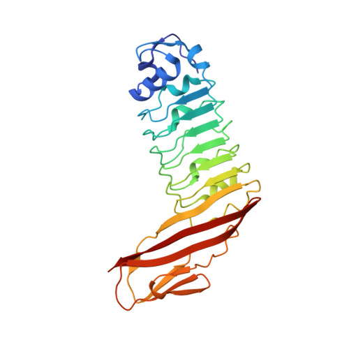
Structure of internalin C from Listeria monocytogenes.

Ooi, A.Hussain, S.Seyedarabi, A.Pickersgill, R.W.

(2006) Acta Crystallogr D Biol Crystallogr 62: 1287-1293

  • DOI: https://doi.org/10.1107/S0907444906026746
  • Primary Citation of Related Structures:  
    1XEU

  • PubMed Abstract: 

    The crystal structure of internalin C (InlC) from Listeria monocytogenes has been determined at 2.0 A resolution. Several observations implicate InlC in infection: inlC has the same transcriptional activator as other virulence genes, it is only present in pathogenic Listeria strains and an inlC deletion mutant is significantly less virulent. While the extended concave receptor-binding surfaces of the leucine-rich repeat (LRR) domains of internalins A and B have aromatic clusters involved in receptor binding, the corresponding surface of InlC is smaller, flatter and more hydrophilic, suggesting that InlC may be involved in weak or transient associations with receptors; this may help explain why no receptor has yet been discovered for InlC. In contrast, the Ig-like domain, to which the LRR domain is fused, has surface aromatics that may be of functional importance, possibly being involved in binding to the surface of the bacteria or in receptor binding.


  • Organizational Affiliation

    School of Biological and Chemical Sciences, Queen Mary, University of London, Mile End Road, London E1 4NS, England.


Macromolecules
Find similar proteins by:  (by identity cutoff)  |  3D Structure
Entity ID: 1
MoleculeChains Sequence LengthOrganismDetailsImage
internalin C263Listeria monocytogenesMutation(s): 0 
Gene Names: inlC
UniProt
Find proteins for P71451 (Listeria monocytogenes serotype 1/2a (strain EGD / Mackaness))
Explore P71451 
Go to UniProtKB:  P71451
Entity Groups  
Sequence Clusters30% Identity50% Identity70% Identity90% Identity95% Identity100% Identity
UniProt GroupP71451
Sequence Annotations
Expand
  • Reference Sequence
Experimental Data & Validation

Experimental Data

  • Method: X-RAY DIFFRACTION
  • Resolution: 2.05 Å
  • R-Value Free: 0.243 
  • R-Value Work: 0.200 
  • R-Value Observed: 0.202 
  • Space Group: P 21 21 2
Unit Cell:
Length ( Å )Angle ( ˚ )
a = 59.327α = 90
b = 177.802β = 90
c = 42.222γ = 90
Software Package:
Software NamePurpose
REFMACrefinement
DENZOdata reduction
SCALEPACKdata scaling
MOLREPphasing

Structure Validation

View Full Validation Report


Entry History 

Deposition Data

Revision History  (Full details and data files)

  • Version 1.0: 2005-08-30
    Type: Initial release
  • Version 1.1: 2008-04-30
    Changes: Version format compliance
  • Version 1.2: 2011-07-13
    Changes: Version format compliance
  • Version 1.3: 2024-02-14
    Changes: Data collection, Database references
  • Version 1.4: 2024-04-03
    Changes: Refinement description