1XB7

X-ray structure of ERRalpha LBD in complex with a PGC-1alpha peptide at 2.5A resolution


Experimental Data Snapshot

  • Method: X-RAY DIFFRACTION
  • Resolution: 2.50 Å
  • R-Value Free: 0.247 
  • R-Value Work: 0.215 
  • R-Value Observed: 0.216 

wwPDB Validation   3D Report Full Report


This is version 1.4 of the entry. See complete history


Literature

Evidence for Ligand-independent Transcriptional Activation of the Human Estrogen-related Receptor {alpha} (ERR{alpha}): CRYSTAL STRUCTURE OF ERR{alpha} LIGAND BINDING DOMAIN IN COMPLEX WITH PEROXISOME PROLIFERATOR-ACTIVATED RECEPTOR COACTIVATOR-1{alpha}

Kallen, J.Schlaeppi, J.M.Bitsch, F.Filipuzzi, I.Schilb, A.Riou, V.Graham, A.Strauss, A.Geiser, M.Fournier, B.

(2004) J Biol Chem 279: 49330-49337

  • DOI: https://doi.org/10.1074/jbc.M407999200
  • Primary Citation of Related Structures:  
    1XB7

  • PubMed Abstract: 

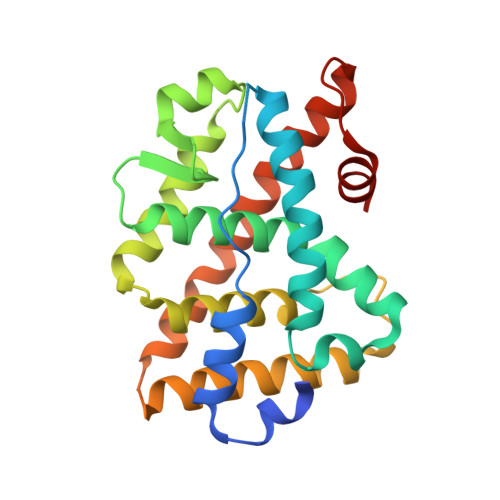
    The crystal structure of the ligand binding domain (LBD) of the estrogen-related receptor alpha (ERRalpha, NR3B1) complexed with a coactivator peptide from peroxisome proliferator-activated receptor coactivator-1alpha (PGC-1alpha) reveals a transcriptionally active conformation in the absence of a ligand. This is the first x-ray structure of ERRalpha LBD, solved to a resolution of 2.5 A, and the first structure of a PGC-1alpha complex. The putative ligand binding pocket (LBP) of ERRalpha is almost completely occupied by side chains, in particular with the bulky side chain of Phe328 (corresponding to Ala272 in ERRgamma and Ala350 in estrogen receptor alpha). Therefore, a ligand of a size equivalent to more than approximately 4 carbon atoms could only bind in the LBP, if ERRalpha would undergo a major conformational change (in particular the ligand would displace H12 from its agonist position). The x-ray structure thus provides strong evidence for ligand-independent transcriptional activation by ERRalpha. The interactions of PGC-1alpha with ERRalpha also reveal for the first time the atomic details of how a coactivator peptide containing an inverted LXXLL motif (namely a LLXYL motif) binds to a LBD. In addition, we show that a PGC-1alpha peptide containing this nuclear box motif from the L3 site binds ERRalpha LBD with a higher affinity than a peptide containing a steroid receptor coactivator-1 motif and that the affinity is further enhanced when all three leucine-rich regions of PGC-1alpha are present.


  • Organizational Affiliation

    Protein Structure Unit, Novartis Institutes for Biomedical Research, Basel, Switzerland. jeorge.kallen@pharma.novartis.com


Macromolecules
Find similar proteins by:  (by identity cutoff)  |  3D Structure
Entity ID: 1
MoleculeChains Sequence LengthOrganismDetailsImage
Steroid hormone receptor ERR1247Homo sapiensMutation(s): 0 
UniProt & NIH Common Fund Data Resources
Find proteins for P11474 (Homo sapiens)
Explore P11474 
Go to UniProtKB:  P11474
PHAROS:  P11474
GTEx:  ENSG00000173153 
Entity Groups  
Sequence Clusters30% Identity50% Identity70% Identity90% Identity95% Identity100% Identity
UniProt GroupP11474
Sequence Annotations
Expand
  • Reference Sequence

Find similar proteins by:  Sequence   |   3D Structure  

Entity ID: 2
MoleculeChains Sequence LengthOrganismDetailsImage
Peroxisome proliferator activated receptor gamma coactivator 1 alphaB [auth P]12N/AMutation(s): 1 
UniProt & NIH Common Fund Data Resources
Find proteins for Q9UBK2 (Homo sapiens)
Explore Q9UBK2 
Go to UniProtKB:  Q9UBK2
PHAROS:  Q9UBK2
GTEx:  ENSG00000109819 
Entity Groups  
Sequence Clusters30% Identity50% Identity70% Identity90% Identity95% Identity100% Identity
UniProt GroupQ9UBK2
Sequence Annotations
Expand
  • Reference Sequence
Small Molecules
Ligands 1 Unique
IDChains Name / Formula / InChI Key2D Diagram3D Interactions
IOD
Query on IOD

Download Ideal Coordinates CCD File 
C [auth P]IODIDE ION
I
XMBWDFGMSWQBCA-UHFFFAOYSA-M
Experimental Data & Validation

Experimental Data

  • Method: X-RAY DIFFRACTION
  • Resolution: 2.50 Å
  • R-Value Free: 0.247 
  • R-Value Work: 0.215 
  • R-Value Observed: 0.216 
  • Space Group: P 65 2 2
Unit Cell:
Length ( Å )Angle ( ˚ )
a = 109.999α = 90
b = 109.999β = 90
c = 104.671γ = 120
Software Package:
Software NamePurpose
REFMACrefinement
DENZOdata reduction
SCALEPACKdata scaling
MOLREPphasing

Structure Validation

View Full Validation Report


Entry History 

Deposition Data

Revision History  (Full details and data files)

  • Version 1.0: 2004-09-14
    Type: Initial release
  • Version 1.1: 2008-04-30
    Changes: Version format compliance
  • Version 1.2: 2011-07-13
    Changes: Version format compliance
  • Version 1.3: 2021-11-10
    Changes: Database references, Derived calculations
  • Version 1.4: 2023-10-25
    Changes: Data collection, Refinement description