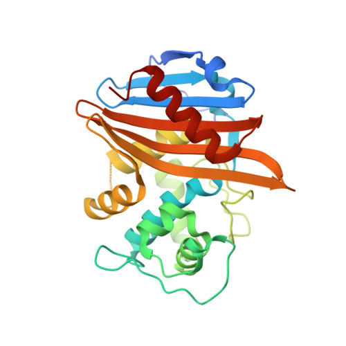
Crystal structures of the Apo and penicillin-acylated forms of the BlaR1 beta-lactam sensor of Staphylococcus aureus.
Wilke, M.S., Hills, T.L., Zhang, H.Z., Chambers, H.F., Strynadka, N.C.(2004) J Biol Chem 279: 47278-47287
- PubMed: 15322076
- DOI: https://doi.org/10.1074/jbc.M407054200
- Primary Citation of Related Structures:
1XA1, 1XA7 - PubMed Abstract:
Staphylococcus aureus is among the most prevalent and antibiotic-resistant of pathogenic bacteria. The resistance of S. aureus to prototypal beta-lactam antibiotics is conferred by two mechanisms: (i) secretion of hydrolytic beta-lactamase enzymes and (ii) production of beta-lactam-insensitive penicillin-binding proteins (PBP2a). Despite their distinct modes of resistance, expression of these proteins is controlled by similar regulation systems, including a repressor (BlaI/MecI) and a multidomain transmembrane receptor (BlaR1/MecR1). Resistance is triggered in response to a covalent binding event between a beta-lactam antibiotic and the extracellular sensor domain of BlaR1/MecR1 by transduction of the binding signal to an intracellular protease domain capable of repressor inactivation. This study describes the first crystal structures of the sensor domain of BlaR1 (BlaRS) from S. aureus in both the apo and penicillin-acylated forms. The structures show that the sensor domain resembles the beta-lactam-hydrolyzing class D beta-lactamases, but is rendered a penicillin-binding protein due to the formation of a very stable acyl-enzyme. Surprisingly, conformational changes upon penicillin binding were not observed in our structures, supporting the hypothesis that transduction of the antibiotic-binding signal into the cytosol is mediated by additional intramolecular interactions of the sensor domain with an adjacent extracellular loop in BlaR1.
Organizational Affiliation:
Department of Biochemistry and Molecular Biology, University of British Columbia, Vancouver, British Columbia V6T 1Z3, Canada.