1X9H

Crystal structure of phosphoglucose/phosphomannose isomerase from Pyrobaculum aerophilum in complex with fructose 6-phosphate


Experimental Data Snapshot

  • Method: X-RAY DIFFRACTION
  • Resolution: 1.50 Å
  • R-Value Free: 0.180 
  • R-Value Work: 0.152 
  • R-Value Observed: 0.153 

wwPDB Validation   3D Report Full Report


Ligand Structure Quality Assessment 


This is version 1.5 of the entry. See complete history


Literature

Structural basis for phosphomannose isomerase activity in phosphoglucose isomerase from Pyrobaculum aerophilum: a subtle difference between distantly related enzymes.

Swan, M.K.Hansen, T.Schoenheit, P.Davies, C.

(2004) Biochemistry 43: 14088-14095

  • DOI: https://doi.org/10.1021/bi048608y
  • Primary Citation of Related Structures:  
    1X9H, 1X9I

  • PubMed Abstract: 

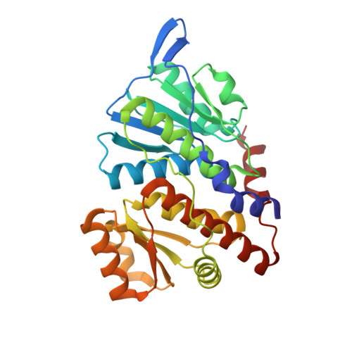
    The crystal structure of a dual-specificity phosphoglucose/phosphomannose isomerase from the crenarchaeon Pyrobaculum aerophilum (PaPGI/PMI) has been determined in complex with glucose 6-phosphate at 1.16 A resolution and with fructose 6-phosphate at 1.5 A resolution. Subsequent modeling of mannose 6-phosphate (M6P) into the active site of the enzyme shows that the PMI activity of this enzyme may be due to the additional space imparted by a threonine. In PGIs from bacterial and eukaryotic sources, which cannot use M6P as a substrate, the equivalent residue is a glutamine. The increased space may permit rotation of the C2-C3 bond in M6P to facilitate abstraction of a proton from C2 by Glu203 and, after a further C2-C3 rotation of the resulting cis-enediolate, re-donation of a proton to C1 by the same residue. A proline residue (in place of a glycine in PGI) may also promote PMI activity by positioning the C1-O1 region of M6P. Thus, the PMI reaction in PaPGI/PMI probably uses a cis-enediol mechanism of catalysis, and this activity appears to arise from a subtle difference in the architecture of the enzyme, compared to bacterial and eukaryotic PGIs.


  • Organizational Affiliation

    Department of Biochemistry and Molecular Biology, Medical University of South Carolina, Charleston, South Carolina 29425, USA.


Macromolecules
Find similar proteins by:  (by identity cutoff)  |  3D Structure
Entity ID: 1
MoleculeChains Sequence LengthOrganismDetailsImage
glucose-6-phosphate isomerase
A, B
302Pyrobaculum aerophilumMutation(s): 0 
Gene Names: PAE1610
EC: 5.3.1.9 (PDB Primary Data), 5.3.1.8 (PDB Primary Data)
UniProt
Find proteins for Q8ZWV0 (Pyrobaculum aerophilum (strain ATCC 51768 / DSM 7523 / JCM 9630 / CIP 104966 / NBRC 100827 / IM2))
Explore Q8ZWV0 
Go to UniProtKB:  Q8ZWV0
Entity Groups  
Sequence Clusters30% Identity50% Identity70% Identity90% Identity95% Identity100% Identity
UniProt GroupQ8ZWV0
Sequence Annotations
Expand
  • Reference Sequence
Small Molecules
Ligands 3 Unique
IDChains Name / Formula / InChI Key2D Diagram3D Interactions
F6R
Query on F6R

Download Ideal Coordinates CCD File 
F [auth A],
L [auth B]
FRUCTOSE -6-PHOSPHATE
C6 H13 O9 P
GSXOAOHZAIYLCY-HSUXUTPPSA-N
SO4
Query on SO4

Download Ideal Coordinates CCD File 
C [auth A],
D [auth A],
E [auth A],
J [auth B],
K [auth B]
SULFATE ION
O4 S
QAOWNCQODCNURD-UHFFFAOYSA-L
GOL
Query on GOL

Download Ideal Coordinates CCD File 
G [auth A],
H [auth A],
I [auth A],
M [auth B]
GLYCEROL
C3 H8 O3
PEDCQBHIVMGVHV-UHFFFAOYSA-N
Experimental Data & Validation

Experimental Data

  • Method: X-RAY DIFFRACTION
  • Resolution: 1.50 Å
  • R-Value Free: 0.180 
  • R-Value Work: 0.152 
  • R-Value Observed: 0.153 
  • Space Group: P 1 21 1
Unit Cell:
Length ( Å )Angle ( ˚ )
a = 55.2α = 90
b = 100.8β = 113.4
c = 55.9γ = 90
Software Package:
Software NamePurpose
REFMACrefinement
MAR345data collection
SCALEPACKdata scaling

Structure Validation

View Full Validation Report


Ligand Structure Quality Assessment 


Entry History 

Deposition Data

Revision History  (Full details and data files)

  • Version 1.0: 2004-12-07
    Type: Initial release
  • Version 1.1: 2008-04-30
    Changes: Version format compliance
  • Version 1.2: 2011-07-13
    Changes: Non-polymer description, Version format compliance
  • Version 1.3: 2017-10-11
    Changes: Refinement description
  • Version 1.4: 2018-01-24
    Changes: Structure summary
  • Version 1.5: 2023-08-23
    Changes: Data collection, Database references, Derived calculations, Refinement description